Difference between revisions of "Prostate cancer staging"
Jump to navigation
Jump to search
(create) |
(→Tumour: split out EPE from main prostate ca page) |
||
| Line 7: | Line 7: | ||
===Tumour=== | ===Tumour=== | ||
====Extraprostatic extension==== | |||
:Abbreviated ''EPE''. | |||
=====General===== | |||
*Extraprostatic extension (EPE) is difficult to assess in prostatectomy specimens.<ref name=pmid20802467>{{Cite journal | last1 = Magi-Galluzzi | first1 = C. | last2 = Evans | first2 = AJ. | last3 = Delahunt | first3 = B. | last4 = Epstein | first4 = JI. | last5 = Griffiths | first5 = DF. | last6 = van der Kwast | first6 = TH. | last7 = Montironi | first7 = R. | last8 = Wheeler | first8 = TM. | last9 = Srigley | first9 = JR. | title = International Society of Urological Pathology (ISUP) Consensus Conference on Handling and Staging of Radical Prostatectomy Specimens. Working group 3: extraprostatic extension, lymphovascular invasion and locally advanced disease. | journal = Mod Pathol | volume = 24 | issue = 1 | pages = 26-38 | month = Jan | year = 2011 | doi = 10.1038/modpathol.2010.158 | PMID = 20802467 }}</ref> | |||
**The prostate does NOT have a well defined capsule. | |||
***Intraobserver agreement for EPE is fair-moderate and lower than for the surgical margin.<ref name=pmid18708939>{{Cite journal | last1 = Evans | first1 = AJ. | last2 = Henry | first2 = PC. | last3 = Van der Kwast | first3 = TH. | last4 = Tkachuk | first4 = DC. | last5 = Watson | first5 = K. | last6 = Lockwood | first6 = GA. | last7 = Fleshner | first7 = NE. | last8 = Cheung | first8 = C. | last9 = Belanger | first9 = EC. | last10 = Amin | first10 = MB. | last11 = Boccon-Gibod | first11 = L. | last12 = Bostwick | first12 = DG. | last13 = Egevad | first13 = L. | last14 = Epstein | first14 = JI. | last15 = Grignon | first15 = DJ. | last16 = Jones | first16 = EC. | last17 = Montironi | first17 = R. | last18 = Moussa | first18 = M. | last19 = Sweet | first19 = JM. | last20 = Trpkov | first20 = K. | last21 = Wheeler | first21 = TM. | last22 = Srigley | first22 = JR. | title = Interobserver variability between expert urologic pathologists for extraprostatic extension and surgical margin status in radical prostatectomy specimens. | journal = Am J Surg Pathol | volume = 32 | issue = 10 | pages = 1503-12 | month = Oct | year = 2008 | doi = 10.1097/PAS.0b013e31817fb3a0 | PMID = 18708939 }}</ref> | |||
*EPE, typically, upstages tumours from T2x to T3a. | |||
=====Prostatectomy specimens===== | |||
EPE is present in a prostatectomy if there is either: | |||
#A "significant bulge" in the contour of the prostate at low power ''and'' no fibromuscular tissue surrounding the malignant cells. | |||
#Malignant cells directly adjacent to peri-prostatic adipose tissue. | |||
Note: | |||
*The apex of the prostate gland may have some skeletal muscle. Thus, it is difficult to define extension at this site. EPE is not called at the apex by some pathologists; however, it is generally believed to exist.<ref name=pmid20802467/> | |||
=====Prostate biopsy===== | |||
EPE is present in prostate biopsy if: | |||
*Tumour touches adipose tissue.<ref name=pmid17707261>{{Cite journal | last1 = Epstein | first1 = JI. | last2 = Srigley | first2 = J. | last3 = Grignon | first3 = D. | last4 = Humphrey | first4 = P. | title = Recommendations for the reporting of prostate carcinoma. | journal = Hum Pathol | volume = 38 | issue = 9 | pages = 1305-9 | month = Sep | year = 2007 | doi = 10.1016/j.humpath.2007.05.015 | PMID = 17707261 }}</ref> | |||
======Images====== | |||
<gallery> | |||
Image: Prostate carcinoma with extraprostatic extension -- intermed mag.jpg | EPE - intermed. mag. | |||
Image: Prostate carcinoma with extraprostatic extension -- high mag.jpg | EPE - high mag. | |||
</gallery> | |||
===Lymph node=== | ===Lymph node=== | ||
Revision as of 03:29, 9 February 2016
The article deals with prostate cancer staging. A general discussion of staging is found in cancer staging.
General
- Important for prognosis and treatment.
TNM staging system
Tumour
Extraprostatic extension
- Abbreviated EPE.
General
- Extraprostatic extension (EPE) is difficult to assess in prostatectomy specimens.[1]
- The prostate does NOT have a well defined capsule.
- Intraobserver agreement for EPE is fair-moderate and lower than for the surgical margin.[2]
- The prostate does NOT have a well defined capsule.
- EPE, typically, upstages tumours from T2x to T3a.
Prostatectomy specimens
EPE is present in a prostatectomy if there is either:
- A "significant bulge" in the contour of the prostate at low power and no fibromuscular tissue surrounding the malignant cells.
- Malignant cells directly adjacent to peri-prostatic adipose tissue.
Note:
- The apex of the prostate gland may have some skeletal muscle. Thus, it is difficult to define extension at this site. EPE is not called at the apex by some pathologists; however, it is generally believed to exist.[1]
Prostate biopsy
EPE is present in prostate biopsy if:
- Tumour touches adipose tissue.[3]
Images
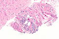
EPE - intermed. mag.
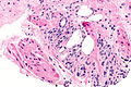
EPE - high mag.


Lymph node
See also
References
- ↑ 1.0 1.1 Magi-Galluzzi, C.; Evans, AJ.; Delahunt, B.; Epstein, JI.; Griffiths, DF.; van der Kwast, TH.; Montironi, R.; Wheeler, TM. et al. (Jan 2011). "International Society of Urological Pathology (ISUP) Consensus Conference on Handling and Staging of Radical Prostatectomy Specimens. Working group 3: extraprostatic extension, lymphovascular invasion and locally advanced disease.". Mod Pathol 24 (1): 26-38. doi:10.1038/modpathol.2010.158. PMID 20802467.
- ↑ Evans, AJ.; Henry, PC.; Van der Kwast, TH.; Tkachuk, DC.; Watson, K.; Lockwood, GA.; Fleshner, NE.; Cheung, C. et al. (Oct 2008). "Interobserver variability between expert urologic pathologists for extraprostatic extension and surgical margin status in radical prostatectomy specimens.". Am J Surg Pathol 32 (10): 1503-12. doi:10.1097/PAS.0b013e31817fb3a0. PMID 18708939.
- ↑ Epstein, JI.; Srigley, J.; Grignon, D.; Humphrey, P. (Sep 2007). "Recommendations for the reporting of prostate carcinoma.". Hum Pathol 38 (9): 1305-9. doi:10.1016/j.humpath.2007.05.015. PMID 17707261.