Difference between revisions of "Acinic cell carcinoma"
(split out) |
(+infobox) |
||
| Line 1: | Line 1: | ||
{{ Infobox diagnosis | |||
| Name = {{PAGENAME}} | |||
| Image = Acinic_cell_carcinoma_-_very_high_mag.jpg | |||
| Width = | |||
| Caption = Acinic cell carcinoma. [[H&E stain]]. | |||
| Micro = "acinic cells" (abundant finely vacuolated cytoplasm with basophilic granules, small nuclei with stippled chromatin), scattered "intercalcated duct type cells" (eosinophilic cytoplasm with moderate amount of cytoplasm and bland nuclei), +/-peri-tumoural lymphocytes. | |||
*+/-glassy extracellular bluish/purple blobs | |||
| Subtypes = oncocytic variant, clear cell variant, papillary cystic variant | |||
| LMDDx = [[oncocytoma of the salivary gland]], [[mucoepidermoid carcinoma]], adenocarcinoma NOS | |||
| Stains = PAS +ve, PASD +ve | |||
| IHC = p63 -ve, S-100 -ve | |||
| EM = [[zymogen granules]] | |||
| Molecular = | |||
| IF = | |||
| Gross = tan or reddish | |||
| Grossing = | |||
| Site = [[salivary gland]] - usu. parotid gland | |||
| Assdx = | |||
| Syndromes = | |||
| Clinicalhx = | |||
| Signs = salivary gland mass | |||
| Symptoms = | |||
| Prevalence = | |||
| Bloodwork = | |||
| Rads = | |||
| Endoscopy = | |||
| Prognosis = | |||
| Other = | |||
| ClinDDx = | |||
}} | |||
'''Acinic cell carcinoma''', abbreviated '''AcCC''', is a rare type of [[salivary gland]] cancer. It is also known as '''acinic cell adenocarcinoma'''. | '''Acinic cell carcinoma''', abbreviated '''AcCC''', is a rare type of [[salivary gland]] cancer. It is also known as '''acinic cell adenocarcinoma'''. | ||
| Line 71: | Line 101: | ||
*S-100 -ve. | *S-100 -ve. | ||
*p63 -ve. | *p63 -ve. | ||
**p63 +ve in mucoepidermoid carcinoma. | **p63 +ve in [[mucoepidermoid carcinoma]]. | ||
There are a bunch of other [[stains]] that are touted to be useful (amylase, anti-chymotrypsin, lactoferrin). Weinreb thinks these are '''not''' helpful.<ref name=IW_10jan2011>IW. 11 January 2011.</ref> | There are a bunch of other [[stains]] that are touted to be useful (amylase, anti-chymotrypsin, lactoferrin). Weinreb thinks these are '''not''' helpful.<ref name=IW_10jan2011>IW. 11 January 2011.</ref> | ||
Revision as of 22:14, 29 July 2013
| Acinic cell carcinoma | |
|---|---|
| Diagnosis in short | |
 Acinic cell carcinoma. H&E stain. | |
|
| |
| LM |
"acinic cells" (abundant finely vacuolated cytoplasm with basophilic granules, small nuclei with stippled chromatin), scattered "intercalcated duct type cells" (eosinophilic cytoplasm with moderate amount of cytoplasm and bland nuclei), +/-peri-tumoural lymphocytes.
|
| Subtypes | oncocytic variant, clear cell variant, papillary cystic variant |
| LM DDx | oncocytoma of the salivary gland, mucoepidermoid carcinoma, adenocarcinoma NOS |
| Stains | PAS +ve, PASD +ve |
| IHC | p63 -ve, S-100 -ve |
| EM | zymogen granules |
| Gross | tan or reddish |
| Site | salivary gland - usu. parotid gland |
|
| |
| Signs | salivary gland mass |
Acinic cell carcinoma, abbreviated AcCC, is a rare type of salivary gland cancer. It is also known as acinic cell adenocarcinoma.
It should not to be confused with pancreatic acinar cell carcinoma.
General
- Malignant neoplasm of salivary gland arising from acinic cells.
- The relative prevalence of the neoplasm in the various salivary gland reflects the abundance of acinic cells: parotid gland (~80%) > minor salivary glands (~17%) > submandibular glands (~3%).
- Affects wide age range -- including children.
- Site affect prognosis (most aggressive to least aggressive): submandibular > parotid > minor salivary.
Gross
- Tan or reddish.
Microscopic
Features:
- Sheets of acinic cells with:
- Abundant finely vacuolated cytoplasm with basophilic granules - key feature.
- Granules may be focal.
- Small nuclei stippled chromatin.
- Abundant finely vacuolated cytoplasm with basophilic granules - key feature.
- Scattered intercalcated duct type cells with:
- Eosinophilic cytoplasm with moderate amount of cytoplasm.
- Bland nuclei with slightly larger than seen in acinic cells.
- +/-Peri-tumoural lymphocytes.
- +/-Glassy extracellular bluish/purple blobs.
Notes:
- Adipose tissue -- present in the salivary glands -- is absent in AcCC.
- May focally resemble thyroid tissue.
- Smaller (characteristic) microvacuoles (unreported in the literature) may be present that have a bubbly appearance and glassy basophilic inclusions.[1]
Memory device:
- AcCC - lots of "C"s - chromatin stipled, cytoplasm generous.
DDx:
- Oncocytoma of the salivary gland.
- Adenocarcinoma not otherwise specified.[2]
Images
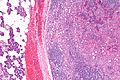
AcCC - intermed. mag. (WC/Nephron)
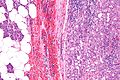
AcCC - high mag. (WC/Nephron)
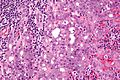
AcCC - very high mag. (WC/Nephron)


www:
- AcCC (surgicalpathologyatlas.com).
- AcCC (brown.edu).
- AcCC (aciniccell.org) - image collection.
Grading
General:
- Not prognostic.
- Done to avoid phone calls from clinician.
Factors Weinreb uses:[1]
- Necrosis.
- Nuclear atypia.
- Perineural invasion.
- Mitoses.
- Infiltrative margin.
- Tumour sclerosis.
Subtypes
- Oncocytic variant - rare.
- Clear cell variant - rare.
- Papillary cystic variant.
Stains
- PAS +ve.
- PAS-D +ve.
IHC
- S-100 -ve.
- p63 -ve.
- p63 +ve in mucoepidermoid carcinoma.
There are a bunch of other stains that are touted to be useful (amylase, anti-chymotrypsin, lactoferrin). Weinreb thinks these are not helpful.[1]
EM
See also
References
- ↑ 1.0 1.1 1.2 IW. 11 January 2011.
- ↑ Ihrler, S.; Blasenbreu-Vogt, S.; Sendelhofert, A.; Lang, S.; Zietz, C.; Löhrs, U. (2002). "Differential diagnosis of salivary acinic cell carcinoma and adenocarcinoma (NOS). A comparison of (immuno-)histochemical markers.". Pathol Res Pract 198 (12): 777-83. PMID 12608654.
- ↑ Sun, Y.; Wasserman, PG. (Feb 2004). "Acinar cell carcinoma arising in the stomach: a case report with literature review.". Hum Pathol 35 (2): 263-5. PMID 14991547.