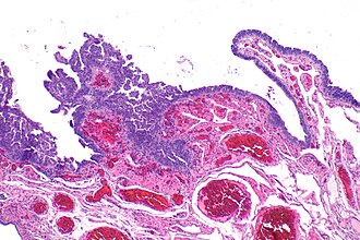
Serous tubal intraepithelial carcinoma
Jump to navigation
Jump to search
| Serous tubal intraepithelial carcinoma | |
|---|---|
| Diagnosis in short | |
 Micrograph showing serous tubal intraepithelial carcinoma. H&E stain. | |
|
| |
| LM | three of the following five: (1) atypical chromatin pattern, (2) nuclear enlargement, (3) nuclear pleomorphism, (4) nuclear moulding, (5) loss of nuclear polarity or epithelial stratification |
| IHC | p16 +ve, p53 +ve, Ki-67 increased |
| Molecular | BRCA1 mutation or BRCA2 mutation |
| Gross | not apparent on gross |
| Site | fallopian tubes |
|
| |
| Symptoms | none |
| Prevalence | uncommon |
Serous tubal intraepithelial carcinoma, abbreviated STIC[1] (pronounced stick), is considered to be the precursor of serous carcinoma.
It is also known as tubal intraepithelial carcinoma.
General
- Considered the precursor lesion for tubal serous carcinoma.[2]
- Seen in ~6% of prophylactic salpingo-oophorectomies in BRCA-mutation carriers.[3]
Gross
- Not apparent on gross.
- Usually at the fimbriated end of the tube.
Microscopic
Features:[4]
- Discrete papillary growth - low power.
- Formal criteria - need 3 or more:
- Atypical chromatin pattern.
- Nuclear enlargement.
- Nuclear pleomorphism.
- Nuclear moulding.
- Loss of nuclear polarity or epithelial stratification.
Notes:
- At low power STIC is usually tall cells that are too blue.
- Cilia suggest benign.
DDx:
- Atypical tubal lesion (STIL or tubal intraepithelial lesions in transition (TILT)[5]) - lack proliferation.[citation needed]
- Serous carcinoma of the fallopian tube.
Images
www
- STIC (nature.com).[4]
- STIC - schematic (nih.gov).[6]
- STIC and STIL with p53 stain (nih.gov).[6]
- STIC - p53 stain (nih.gov).[6]
- Small STIC - H&E and p53 (nih.gov).[7]
- STIC - high mag. (ovariancancerprevention.org).[8]
- STIC & benign - high mag. (ovariancancerprevention.org).
IHC
Features:[1]
- p53 +ve.
- Ki-67 increased ~10%. (???)
- p16 +ve.[4]
Images
See also
References
- ↑ 1.0 1.1 Visvanathan, K.; Vang, R.; Shaw, P.; Gross, A.; Soslow, R.; Parkash, V.; Shih, IeM.; Kurman, RJ. (Dec 2011). "Diagnosis of serous tubal intraepithelial carcinoma based on morphologic and immunohistochemical features: a reproducibility study.". Am J Surg Pathol 35 (12): 1766-75. doi:10.1097/PAS.0b013e31822f58bc. PMID 21989347.
- ↑ Lee, Y.; Miron, A.; Drapkin, R.; Nucci, MR.; Medeiros, F.; Saleemuddin, A.; Garber, J.; Birch, C. et al. (Jan 2007). "A candidate precursor to serous carcinoma that originates in the distal fallopian tube.". J Pathol 211 (1): 26-35. doi:10.1002/path.2091. PMID 17117391.
- ↑ Mingels MJ, Roelofsen T, van der Laak JA, et al. (October 2012). "Tubal epithelial lesions in salpingo-oophorectomy specimens of BRCA-mutation carriers and controls". Gynecol. Oncol. 127 (1): 88–93. doi:10.1016/j.ygyno.2012.06.015. PMID 22710074.
- ↑ 4.0 4.1 4.2 Sehdev, AS.; Kurman, RJ.; Kuhn, E.; Shih, IeM. (Jun 2010). "Serous tubal intraepithelial carcinoma upregulates markers associated with high-grade serous carcinomas including Rsf-1 (HBXAP), cyclin E and fatty acid synthase.". Mod Pathol 23 (6): 844-55. doi:10.1038/modpathol.2010.60. PMID 20228782.
- ↑ Nishida N, Murakami F, Higaki K (June 2016). "Detection of serous precursor lesions in resected fallopian tubes from patients with benign diseases and a relatively low risk for ovarian cancer". Pathol Int 66 (6): 337–42. doi:10.1111/pin.12419. PMID 27250113.
- ↑ 6.0 6.1 6.2 Kurman, RJ.; Shih, IeM. (Jul 2011). "Molecular pathogenesis and extraovarian origin of epithelial ovarian cancer--shifting the paradigm.". Hum Pathol 42 (7): 918-31. doi:10.1016/j.humpath.2011.03.003. PMID 21683865.
- ↑ Gross, AL.; Kurman, RJ.; Vang, R.; Shih, IeM.; Visvanathan, K. (2010). "Precursor lesions of high-grade serous ovarian carcinoma: morphological and molecular characteristics.". J Oncol 2010: 126295. doi:10.1155/2010/126295. PMID 20445756.
- ↑ URL: http://www.ovariancancerprevention.org/?page_id=191. Accessed on: 13 May 2014.


