Subependymal giant cell astrocytoma
| Subependymal giant cell astrocytoma | |
|---|---|
| Diagnosis in short | |
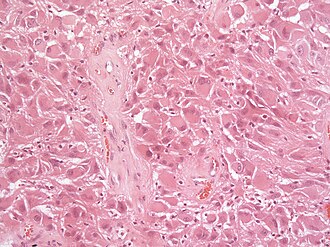
 Subependymal giant cell astrocytoma H&E stain. | |
|
| |
| Synonyms | SEGA |
| LM DDx | ganglioglioma, pleomorphic xanthoastrocytoma, glioblastoma |
| IHC | GFAP +ve |
| Site | brain - usu. wall of ventricles |
|
| |
| Prevalence | rare - esp. in young adults |
| Prognosis | good (WHO Grade I) |
Subependymal giant cell astrocytoma, abbreviated SEGA, is a low-grade astrocytoma associated with tuberous sclerosis complex.
General
- Associated with tuberous sclerosis complex (TSC).[1]
- 6-14% of all TSC patients will develop a SEGA.
- Sporadic examples of SEGA may represent undetected TSC patients (i.e., low-level somatic mosaicism)[2].
- Associated with epilepsy.
- WHO Grade I.
Gross/radiology
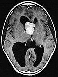
- Well-demarcated.
- Often projecting into a ventricle.
- May be calcified
- Circumscribed tumour.
Microscopic
- Giant cells with nuclear atypia ("bizarre cells", "ganglioid cells").
- Glassy eosinophilic cytoplasm.
- Elongated cells in a fibrillary background.
- Abundant mast cells.[5]
- Lymphocytic infiltrates.
- Endothelial proliferations and/or necrosis are not a sign of malignancy.
Images
www:
IHC
- GFAP +ve. (50%)
- Vimentin +ve. (100%)
- S100 +ve. (100%)
- Neurofilament +/-ve (ganglionic component).
- Synaptophysin +/-ve (ganglionic component)..
- TTF-1 (7 out of 7).[7]
- Olig2-ve.[8]
- MIB-1 usu. low (1-5%).
See also
References
- ↑ Grajkowska, W.; Kotulska, K.; Jurkiewicz, E.; Roszkowski, M.; Daszkiewicz, P.; Jóźwiak, S.; Matyja, E. (2011). "Subependymal giant cell astrocytomas with atypical histological features mimicking malignant gliomas.". Folia Neuropathol 49 (1): 39-46. PMID 21455842.
- ↑ Overwater, IE.; Swenker, R.; van der Ende, EL.; Hanemaayer, KB.; Hoogeveen-Westerveld, M.; van Eeghen, AM.; Lequin, MH.; van den Ouweland, AM. et al. (12 2016). "Genotype and brain pathology phenotype in children with tuberous sclerosis complex.". Eur J Hum Genet 24 (12): 1688-1695. doi:10.1038/ejhg.2016.85. PMID 27406250.
- ↑ 3.0 3.1 URL: http://path.upmc.edu/cases/case179.html. Accessed on: 29 July 2011.
- ↑ 4.0 4.1 Taraszewska, A.; Kroh, H.; Majchrowski, A. (1997). "Subependymal giant cell astrocytoma: clinical, histologic and immunohistochemical characteristic of 3 cases.". Folia Neuropathol 35 (3): 181-6. PMID 9595853.
- ↑ 5.0 5.1 URL: http://path.upmc.edu/cases/case179/micro.html. Accessed on: 8 January 2012.
- ↑ Hirose, T.; Scheithauer, BW.; Lopes, MB.; Gerber, HA.; Altermatt, HJ.; Hukee, MJ.; VandenBerg, SR.; Charlesworth, JC. (1995). "Tuber and subependymal giant cell astrocytoma associated with tuberous sclerosis: an immunohistochemical, ultrastructural, and immunoelectron and microscopic study.". Acta Neuropathol 90 (4): 387-99. PMID 8546029.
- ↑ Hewer, E.; Vajtai, I.. "Consistent nuclear expression of thyroid transcription factor 1 in subependymal giant cell astrocytomas suggests lineage-restricted histogenesis.". Clin Neuropathol 34 (3): 128-31. doi:10.5414/NP300818. PMID 25669749.
- ↑ Overwater, IE.; Swenker, R.; van der Ende, EL.; Hanemaayer, KB.; Hoogeveen-Westerveld, M.; van Eeghen, AM.; Lequin, MH.; van den Ouweland, AM. et al. (12 2016). "Genotype and brain pathology phenotype in children with tuberous sclerosis complex.". Eur J Hum Genet 24 (12): 1688-1695. doi:10.1038/ejhg.2016.85. PMID 27406250.


