Retinoblastoma
Jump to navigation
Jump to search
| Retinoblastoma | |
|---|---|
| Diagnosis in short | |
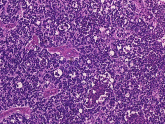
 Retinoblastoma. (WC) | |
|
| |
| LM | small round cell tumour |
| Site | eye |
|
| |
| Prevalence | rare |
Retinoblastoma is malignant tumour the of the eye.
General
Gross
- White, solid.
- Patterns:
- Endophytic - grow into the vitreous cavity.
- Exophytic - grow toward choroid.
- Mixed - components of endophytic and exophytic.
Note:
- Tumour is extremely friable.
Image
Microscopic
Features:
- Small round cell tumour:
- Scant cytoplasm.
- Flexner-Wintersteiner rosette - key feature.
- Rosette with empty centre (donut hole).[2]
- +/-Homer-Wright rosette.[3]
- Circular rosette with neuropil at the centre.[2]
- Mitoses - common.
- +/-Necrosis.
- +/-Calcification.
DDx:
- Retinocytoma (retinoma) - benign counterpart of retinoblastoma.
Notes:
- DDx of Flexner-Wintersteiner rosette includes:
- Pineoblastoma.
- Medulloepithelioma.
