Glioblastoma
| Glioblastoma | |
|---|---|
| Diagnosis in short | |
|
Template:Px Glioblastoma. H&E stain. | |
|
| |
| LM | astrocytic differentiation, nuclear atypia, necrosis, microvascular proliferation, +/-pseudopalisading necrosis |
| Subtypes | gliosarcoma |
| LM DDx | anaplastic astrocytoma |
| IHC | GFAP +ve, IDH-1 -ve/+ve |
| Site | brain, spinal cord |
|
| |
| Radiology | intra-axial |
| Prognosis | very poor |
| Clin. DDx | metastatic brain tumours |
Glioblastoma a very common malignant primary brain tumour in adults. It has a very poor prognosis.
It was previously known as glioblastoma multiforme, abbreviated GBM.
General
- Median survival is measured in months.[1]
- Only about 5% can expect to survive more than three years.[2]
Microscopic
Features:
- Astrocytic tumour with:
- Nuclear atypia.
- Necrosis.
- Endothelial proliferation (AKA microvascular proliferation).
- +/-"Pseudopalisading necrosis" - tumour cells lined-up like a picket fence around necrotic areas.
Images

Core features of GBM: Necrosis, MVP and mitoses (WC/jensflorian)
Anaplastic cells in GBM (WC/jensflorian)
Endothelial proliferations in GBM (WC/jensflorian)
Mitotic activity in GBM (WC/jensflorian)
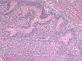
Pseudopalisading necrosis in GBM (WC/jensflorian)
- Glioblastoma brain infiltration zone.jpg
Diffuse brain infiltration in GBM (WC/jensflorian)
- Glioma vessels.jpg
MVP adjacent to tumor infiltration border in GBM (WC/jensflorian)
- Glioblastoma - low mag.jpg
GBM - low mag. (WC)
- Glioblastoma - intermed mag.jpg
GBM juxtaposed with near normal white matter - intermed. mag. (WC)
- Glioblastoma - high mag.jpg
GBM juxtaposed with near normal white matter - high mag. (WC)
- Glioblastoma - very high mag.jpg
GBM - very high mag. (WC)
- Glioblastoma with extreme nuclear enlargement - high mag.jpg
GBM - high mag. (WC)
- Glioblastoma with extreme nuclear enlargement - very high mag.jpg
Extreme nuclear enlargement in a GBM - very high mag. (WC)
- Giant cell glioblastoma HE X200.jpg
Giant cell glioblastoma (WC/jensflorian)
- Adenoid glioblastoma HE.jpg
Adenoid growth pattern in GBM (WC/jensflorian)
- AFIP-00405573-Glioblastoma-Micro.jpg
Adenoid growth pattern in GBM (AFIP)
- Epitheloid glioblastoma HE.jpg
Epitheloid glioblastoma (WC/jensflorian)
- GBM PNET HE x10.jpg
GBM with PNET component (WC/jensflorian)
- Glioblastoma PNET component.jpg
GBM with PNET component (WC/jensflorian)
- Glioblastoma oligodendroglial features.jpg
GBM with oligodendroglial component (WC/jensflorian)
- Glioblastoma pleomoprhism HE.jpg
Nuclear pleomorphism in with oligodendroglial component (WC/jensflorian)
- Glioblastoma granular cell astrocytoma component.jpg
GBM with granular cell component (WC/jensflorian)
- Glioblastoma ependymal features.jpg
Glioblastoma with ependymal-like growth pattern (WC/jensflorian)
- AFIP00405522M-GLIOBLASTOMA ARISING IN ASTROCYTOMA.jpg
Spinal cord GBM (AFIP)
- GBM mimicking melanoma.jpg
Glioblastoma mimicking a (amelanotic) melanoma (WC/jensflorian)
- GBM layers.jpg
Resection borders in a recurrent GBM (WC/jensflorian)
Radiation changes in a recurrent GBM (WC/jensflorian)
GFAP immunostaining in GBM (WC/Marvin 101)
- Wilms tumor protein wt1 immunohistocehmistry glioblastoma.JPG
WT1 immunostaining in GBM (WC/jensflorian)
- Glioblastoma P53.jpg
GBM with strong p53 immunreactivity (WC/jensflorian)


www:
- Microvascular proliferation in a GBM (ouhsc.edu).
- Pseudopalisading necrosis in GBM (aacrjournals.org).

IHC
- GFAP +ve (cytoplasm).
- IDH-1 -ve.
- +ve if developed from lower grade astrocytoma. (???)
See also
References
- ↑ Jubelirer, SJ.. "A review of the treatment and survival rates of 138 patients with glioblastoma multiforme.". W V Med J 92 (4): 186-90. PMID 8772403.
- ↑ Krex, D.; Klink, B.; Hartmann, C.; von Deimling, A.; Pietsch, T.; Simon, M.; Sabel, M.; Steinbach, JP. et al. (Oct 2007). "Long-term survival with glioblastoma multiforme.". Brain 130 (Pt 10): 2596-606. doi:10.1093/brain/awm204. PMID 17785346.