Urinary bladder xanthoma
Jump to navigation
Jump to search
| Urinary bladder xanthoma | |
|---|---|
| Diagnosis in short | |
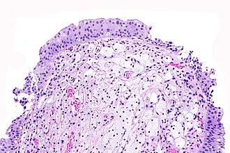
 Urinary bladder xanthoma. H&E stain. | |
|
| |
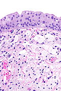
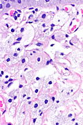
| LM | aggregate of foamy histiocytes in the lamina propria, no significant nuclear atypia |
| LM DDx | signet ring cell carcinoma, granular cell tumour, malakoplakia, xanthogranulomatous cystitis, benign urothelium with mild atypia |
| IHC | CD68 +ve, S-100 -ve |
| Site | urinary bladder (elsewhere) |
|
| |
| Associated Dx | lipid disorders (hyperlipidemia) |
| Signs | +/-hematuria |
| Symptoms | irritation |
| Prevalence | very rare |
| Endoscopy | patch or plaque, yellow, ill-defined border |
| Prognosis | benign |
Urinary bladder xanthoma, also xanthoma of the urinary bladder and bladder xanthoma, is a rare benign lesion of the bladder that occasionally may be clinically suspicious for malignancy.[1]
General
- Rare.
- Like other xanthomas may be associated with abnormal lipid profile.
Presentation:[2]
- Hematuria.
- Irritation.
Gross
Features:[2]
Microscopic
Features:
- Aggregate of foamy histiocytes in the lamina propria.[2]
- No significant nuclear atypia.
DDx:[2]
- Signet ring cell carcinoma.
- Granular cell tumour.
- Malakoplakia.
- Xanthogranulomatous cystitis.
- Mild urothelial atypia (esp. large umbrella cells) in normal urothelium.
Images
IHC
- CD68 +ve.
- S-100 -ve
- Positive in granular cell tumour.
Sign out
Urinary Bladder Lesion, Transurethral Excision:
- Xanthoma, see comment.
- Negative for carcinoma in situ and negative for invasive malignancy.

COMMENT:
These lesions may be associated metabolic abnormalities. A lipid profile
should be considered.
See also
References
- ↑ Hassouna, H.; Broome, JD.; Swalem, K.; Manikandan, R. (2014). "Xanthoma of the urinary bladder: a rare benign condition which may be mistaken for malignancy.". BMJ Case Rep 2014. doi:10.1136/bcr-2014-203836. PMID 24810457.
- ↑ 2.0 2.1 2.2 2.3 Yu, DC.; Patel, P.; Bonert, M.; Carlson, K.; Yilmaz, A.; Paner, G.; Magi-Galluzzi, C.; Lopez-Beltran, A. et al. (Aug 2015). "Urinary bladder xanthoma: a multi-institutional series of 17 cases.". Histopathology 67 (2): 255-61. doi:10.1111/his.12647. PMID 25580863.


