Bone
Jump to navigation
Jump to search
Bone is a scaffold it bears weight and occasionally gets infected.
Tumours often spread to bone and occasionally arise in bone. Bone tumours are dealt with in the bone tumours article.
Normal bone
Bone
- Normal bone has osteocytes.
- If the osteocytes are missing... the bone is dead.
- Osteoblasts - make bone.
- Osteoclasts - destroy bone.
Memory device: 'b' before 'c'.
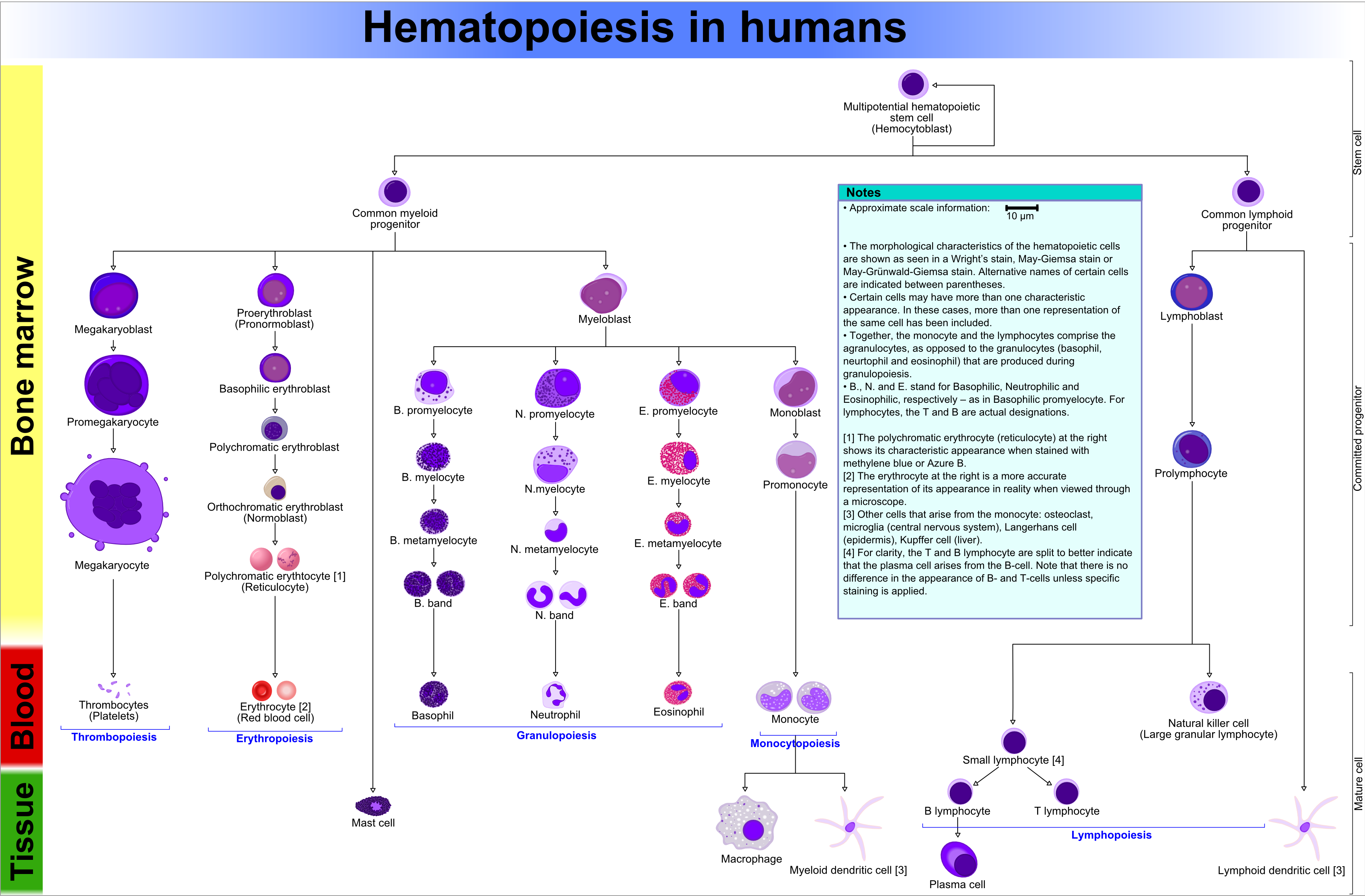
Bone marrow
Main article: Haematopoiesis
- Fat content (%) ~= age (in years)[1]
- e.g. 60 year old will have 60% fatty replacement.
- Should see three cell lines.
- The cell lines:[2]
- Erythroid (red cells),
- Myeloid (white blood cells),
- Megakaryocytic (platelets).
- The cell lines:[2]
Note: Lymphocytes are considered separately and typically spared in bone marrow failure.[3]
Identifying the lines:[4]
- Megakaryocytes:
- Big cells ~ 3x the size of a RBC.
- Normoblasts (RBC precursors):
- Hyperchromatic, i.e. blue, nucleus.
- Myeloid line:
- Granules.
- Reniform nucleus, i.e. kidney bean shaped nucleus.
Images:
Organization
- Mature hematopoeitic cells at the centre (distant from bone).
- Immature hematopoeitic cells adjacent to the bone.
Benign variants
Hyperostosis frontalis interna
- Extra-thick frontal bone.[5]
- No clinical significance -- just has to be recognized as a "nothing".
Infections
Osteomyelitis
General
- Hematogenous - often in children.
- Direct entry (skin defect) - adults with diabetes.
Microscopic
- PMNs.
Chronic osteomyelitis
- Plasma cells.
- May be sterile, i.e. no organisms.
Bone tumours
Main article: Bone tumours
This is a big topic. It is dealt with in a separate article.
The bone tumour article covers tumour mimics, e.g. brown cell tumour.
Fractures
Main article: Forensic pathology
This is dealt with in the forensic pathology article.
Others
The following is a collection of weird stuffs.
Myositis ossificans
General
Epidemiology:
- Young people.
- History of trauma - typically.
- Extremities - digits (fingers, toes).
Notes:
- Histologically "worrisome" (for malignancy) - due to high cellularity.[6]
Microscopic
Features:[6]
- High cellularity.
- Low mitotic activity.
- No atypical mitoses.
- No hyperchromasia.
Other features:[7]
- Low power diagnosis:
- Lesion is well-circumscribed.
- Normal muscle is adjacent to the lesion - key feature.
Paget disease of the bone
General
- Benign - unlike Paget disease of the breast.
Classically divided into three phases:[8][9]
- Lytic (predominantly osteoclasts).
- Mixed lytic (osteoclastic) and blastic (osteoblastic).
- Sclerotic (burned-out).
Clinical:
- Elevated ALP.
Microscopic
Features:[8]
- Bone matrix has jigsaw-puzzle like pattern.
- Jigsaw-puzzle pieces each ~ 100-500 micrometres in size (largest dimension).
- Increased osteoclast activity.
- Osteoclast = macrophage that resorbs bone matrix.
Images:
See also
References
- ↑ IAV. 26 Feb 2009.
- ↑ http://emedicine.medscape.com/article/199003-overview
- ↑ http://emedicine.medscape.com/article/199003-overview
- ↑ http://upload.wikimedia.org/wikipedia/commons/6/69/Hematopoiesis_%28human%29_diagram.png
- ↑ URL: http://radiopaedia.org/articles/hyperostosis_frontalis_interna. Accessed on: 29 September 2010.
- ↑ 6.0 6.1 6.2 Humphrey, Peter A; Dehner, Louis P; Pfeifer, John D (2008). The Washington Manual of Surgical Pathology (1st ed.). Lippincott Williams & Wilkins. pp. 607. ISBN 978-0781765275.
- ↑ IAV. 9 December 2010.
- ↑ 8.0 8.1 URL: http://emedicine.medscape.com/article/311688-overview. Accessed on: 25 December 2010.
- ↑ URL: http://radiopaedia.org/articles/paget-disease-of-bone-1. Accessed on: 25 December 2010.