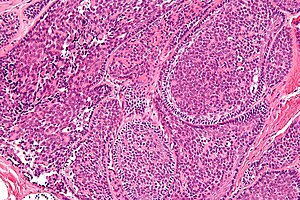
Basal cell adenocarcinoma
Jump to navigation
Jump to search
| Basal cell adenocarcinoma | |
|---|---|
| Diagnosis in short | |
 Basal cell adenocarcinoma. H&E stain. | |
|
| |
| LM | unencapsulated lesion with basal-like cells, tubular component - within basal component, dense hyaline stroma around tumour cells |
| LM DDx | adenoid cystic carcinoma, basal cell adenoma |
| Site | salivary gland - usu. parotid gland |
|
| |
Basal cell adenocarcinoma, abbreviated BCAC, is an uncommon malignant salivary gland tumour.
It should not be confused with basal cell carcinoma, a very common tumour of the skin.
General
- Very rare.
- Malignant.
- Good prognosis.
- May arise from a basal cell adenoma.[1]
Gross
- Usually in the parotid gland ~90% of cases.[1]
Microscopic
Features:
- Lesion is not encapsulated - key feature.
- Basal-like cells:
- Basophilic cells - key feature.
- Usually in nests.
- May be bilayered tubules or trabeculae.
- Large basophilic nucleus.
- Minimal-to-moderate eosinophilic cytoplasm.
- Stromal cells.
- Plump spindle cells without significant nuclear atypia.
- Stromal cell nuclei width ~= diameter RBC.
- Dense hyaline stroma.
- Plump spindle cells without significant nuclear atypia.
- Tubular component.
- Within basal component, may be minimal.
DDx:
- Adenoid cystic carcinoma.
- Basal cell adenoma - encapsulated.
- Basaloid squamous cell carcinoma.
Images
www:
- Basal cell adenocarcinoma - intermed. mag. (webpathology.com).
- Basal cell adenocarcinoma - high mag. (webpathology.com).
IHC
Features:[2]
- CK7 +ve (strong).
- S100 +ve/-ve.
See also
References
- ↑ 1.0 1.1 Muller, S.; Barnes, L. (Dec 1996). "Basal cell adenocarcinoma of the salivary glands. Report of seven cases and review of the literature.". Cancer 78 (12): 2471-7. PMID 8952553.
- ↑ Farrell, T.; Chang, YL. (Oct 2007). "Basal cell adenocarcinoma of minor salivary glands.". Arch Pathol Lab Med 131 (10): 1602-4. doi:10.1043/1543-2165(2007)131[1602:BCAOMS]2.0.CO;2. PMID 17922602.


