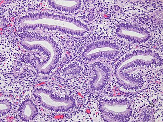
Secretory phase endometrium
Jump to navigation
Jump to search
| Secretory phase endometrium | |
|---|---|
| Diagnosis in short | |
 Early secretory phase. H&E stain. | |
|
| |
| LM | dependent on day post-ovulation - see microscopic |
| LM DDx | endometrial hyperplasia with secretory changes, endometrium with hormonal changes,proliferative phase endometrium |
| Site | endometrium |
|
| |
| Other | normal finding |
Secretory phase endometrium, abbreviated SPE, is a common diagnosis is endometrial specimens.
General
- Secretory phase = luteal phase.
- Gynecologists prefer the ovarian descriptor, i.e. luteal phase; pathologists go by what they see, i.e. Secretions in the (endometrial) glands.
Gross
- Thickened endometrium.
Microscopic
Early secretory phase
Features - post-ovulatory day 1-5:[1]
- Glands: secretory vacuoles.
- First basal to the epithelial nuclei (infranuclear vacuoles).
- Then apical to the epithelial nuclei (supranuclear vacuoles).
- Mitoses may be present - common when vacuoles are subnuclear.
Mid secretory phase
Features - post-ovulatory day 6-8:[1]
- Glands: Mucus in glands.
- Stroma: Edema (empty space around the glands).
Late secretory phase
Features - post-ovulatory day 9-12:[1]
- Stroma:
- Spiral arterioles.
- Predecidual changes -- mnemonic NEW:
- Nucleus central.
- Eosinophilic cytoplasm key feature (may be subtle to the novice).
- Well-defined cell borders.
Premenstrual
- Stroma: neutrophils, scattered lymphocytes, stromal balls ("blue balls"); "stromal condensation".
- Glands: apoptosis at the base of the gland.[2]
Notes:
- Stromal condensation (stromal balls) - premenstrual - stromal cells tightly packed together; nuclei molded together like in small cell tumours.[3]
- Gland-to-stroma ratio is increased in late secretory phase and menstruation.[4]
- Endocervical epithelium (ECE) has a morphology similar to the epithelium of secretory phase endometrium (SPE):
- ECE - grey foamy appearing cytoplasm.
- SPE - eosinophilic cytoplasm.
- Most useful feature to differentiate ECE and SPE is the accompanying stroma.
DDx:
- Endometrial hyperplasia with secretory changes.
- Endometrium with hormonal changes.
- Proliferative phase endometrium - may have some changes of secretory endometrium; <50% of glands have subnuclear vacuoles or <50% of cells in the glands have subnuclear vacuoles.[5]
Images
Sign out
ENDOMETRIUM, BIOPSY:
- SECRETORY PHASE ENDOMETRIUM.
ENDOMETRIUM, CURETTAGE:
- SECRETORY PHASE ENDOMETRIUM.
ENDOMETRIUM, BIOPSY:
- SECRETORY PHASE ENDOMETRIUM, EARLY.
With additional stuff
ENDOMETRIUM, BIOPSY:
- SECRETORY PHASE ENDOMETRIUM.
- SCANT ENDOCERVICAL MUCOSA WITHIN NORMAL LIMITS.
ENDOMETRIUM, BIOPSY:
- SECRETORY PHASE ENDOMETRIUM.
- ENDOCERVICAL MUCOSA AND STRIPPED ENDOCERVICAL EPITHELIUM WITHIN NORMAL LIMITS.
ENDOMETRIUM, BIOPSY:
- SECRETORY PHASE ENDOMETRIUM.
- BENIGN SUPERFICIAL EXOCERVICAL EPITHELIUM.
- SCANT BENIGN ENDOCERVICAL EPITHELIUM.
Evidence of shedding
ENDOMETRIUM, CURETTAGE:
- SECRETORY PHASE ENDOMETRIUM WITH FINDINGS SUGGESTIVE OF SHEDDING (EPITHELIAL
  APOPTOSIS, INFLAMMATORY CELLS - ESPECIALLY NEUTROPHILS).
- BENIGN EXOCERVICAL AND ENDOCERVICAL MUCOSA.
Micro
The sections show endometrium with a normal gland-to-stroma ratio. The glands are mildly dilated, tortuous and have mucous with in them. The glandular epithelium is simple and non-pseudostratified. The stroma is edematous and has a decidual reaction. No mitotic activity is apparent.
See also
References
- ↑ 1.0 1.1 1.2 Tadrous, Paul.J. Diagnostic Criteria Handbook in Histopathology: A Surgical Pathology Vade Mecum (1st ed.). Wiley. pp. 237. ISBN 978-0470519035.
- ↑ Colgan T. 22 June 2009.
- ↑ GAG. 6 Oct 2009.
- ↑ URL: http://www.pathologyoutlines.com/topic/uteruspatternapproach.html. Accessed on: 6 December 2012.
- ↑ Mazur, Michael T.; Kurman, Robert J. (2005). Diagnosis of Endometrial Biopsies and Curettings: A Practical Approach (2nd ed.). Springer. pp. 14. ISBN 978-0387986159.


