Difference between revisions of "Eccrine hidrocystoma"
Jump to navigation
Jump to search
(+omages) |
|||
| Line 52: | Line 52: | ||
==See also== | ==See also== | ||
*[[Eye]]. | *[[Eye]]. | ||
*[[Epidermal inclusion cyst]]. | |||
==References== | ==References== | ||
Revision as of 16:09, 18 January 2017
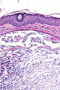
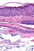
Eccrine hidrocystoma is a benign lesion of the eyelid.
It is occasionally spelled eccrine hydrocystoma.[1]
General
- Benign.
- Eyelid lesion.
Clinical DDx:[1]
- Cystic BCC.
Microsopic
- Cyst lined by a bland bilayer.
- Inner lining cells have:
- Small, round nuclei - usu. basal.
- Moderate pale/greyish cytoplasm.
- No apocrine snouts - flat surface.
- Outer lining cells - spindled.
- May be difficult to see.
- Inner lining cells have:
Note:
- According to the CMAJ, it has the histology of an epidermal inclusion cyst.[1]
DDx:
- Apocrine hidrocystoma - have apocrine snouts; surface is not flat.
- Cystadenoma - has epithelial proliferation.
Image
www
- Eccrine hidrocystoma (nature.com).
- Apocrine hidrocystoma (dermnet.org).
- Apocrine hidrocystoma (flickr.com).
Sign out
SKIN LESION, ADJACENT TO RIGHT EYELID, EXCISION:
- ECCRINE HIDROCYSTOMA.
Micro
The sections show hair bearing skin with a cyst lined by a bland bilayered epithelium. The predominant lining cell has moderate pale grey cytoplasm and a small round nucleus.
The lesion is excised in the plane of section.
See also
References
- ↑ 1.0 1.1 1.2 Adams, SP. (Feb 1999). "Dermacase. Eccrine hydrocystoma.". Can Fam Physician 45: 297, 306. PMC 2328272. PMID 10065300. https://www.ncbi.nlm.nih.gov/pmc/articles/PMC2328272/.
- ↑ Singh, AD.; McCloskey, L.; Parsons, MA.; Slater, DN. (Jan 2005). "Eccrine hidrocystoma of the eyelid.". Eye (Lond) 19 (1): 77-9. doi:10.1038/sj.eye.6701404. PMID 15205675.
- ↑ Busam, Klaus J. (2009). Dermatopathology: A Volume in the Foundations in Diagnostic Pathology Series (1st ed.). Saunders. pp. 314. ISBN 978-0443066542.


