|
|
| Line 275: |
Line 275: |
| ===Diagnosis=== | | ===Diagnosis=== |
| {{hidden|Diagnosis|<center>BORDERLINE SEROUS TUMOR OF THE OVARY WITH MICROINVASION.</center> | | {{hidden|Diagnosis|<center>BORDERLINE SEROUS TUMOR OF THE OVARY WITH MICROINVASION.</center> |
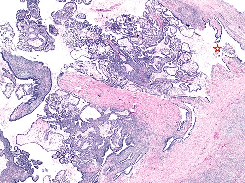
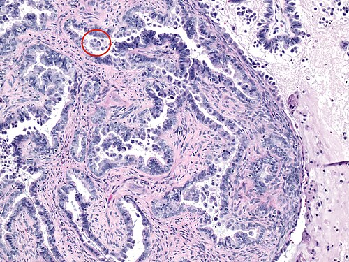
| <br>The characteristic feature of borderline serous tumor is the hierarchical branching of the papillae, where the papillae progressively branch from larger ones to smaller one and finally into tufts of epithelial cells. The cells show only mild to moderate atypia. A cytadenofibroma may have similar proliferation, but by convention should be less than 5% of the tumor. Focal mucinous change may also occur. Papillary clear cell carcinoma simulating borderline tumor will show a WT1-ve, ER-ve, PR-ve immunoprofile in contrast to the positive profile of the serous tumor. Often clusters of cells with abundant eosinophilic cytoplasm are seen at the surface of papillae (star, image 2; oval image 3) or in lymphatic like spaces either as single cells or small papillary clusters. This theoretically represents microinvasion which is variably defined by different authors (<3 or 5 mm or 10mm2). However, recent evidence suggests that the two patterns of invasion have vastly different prognosis. The AEC (abundant eosinophilic cells) are p16 positive and reflect a terminally differentiated senescent phenotype. If invasion is exclusively composed of this cell type, then these do not portend a poor prognosis.(Am J Surg Pathol 2014;38:743–755). These cells are significantly more often seen in BRAF mutated tumors, and are perhaps associated with a better prognosis. (Am J Surg Pathol 2014;38:1603–1611) ( </center>}} | | <br>The characteristic feature of borderline serous tumor is the hierarchical branching of the papillae, where the papillae progressively branch from larger ones to smaller one and finally into tufts of epithelial cells. The cells show only mild to moderate atypia. A cytadenofibroma may have similar proliferation, but by convention should be less than 5% of the tumor. Focal mucinous change may also occur. Papillary clear cell carcinoma simulating borderline tumor will show a WT1-ve, ER-ve, PR-ve immunoprofile in contrast to the positive profile of the serous tumor. Often clusters of cells with abundant eosinophilic cytoplasm are seen at the surface of papillae (star, image 2; oval image 3) or in lymphatic like spaces either as single cells or small papillary clusters. This theoretically represents microinvasion which is variably defined by different authors (<3 or 5 mm or 10mm2). However, recent evidence suggests that the two patterns of invasion have vastly different prognosis. The AEC (abundant eosinophilic cells) are p16 positive and reflect a terminally differentiated senescent phenotype. If invasion is exclusively composed of this cell type, then these do not portend a poor prognosis.<ref name=pmid24441661>{{Cite journal | last1 = Maniar | first1 = KP. | last2 = Wang | first2 = Y. | last3 = Visvanathan | first3 = K. | last4 = Shih | first4 = IeM. | last5 = Kurman | first5 = RJ. | title = Evaluation of microinvasion and lymph node involvement in ovarian serous borderline/atypical proliferative serous tumors: a morphologic and immunohistochemical analysis of 37 cases. | journal = Am J Surg Pathol | volume = 38 | issue = 6 | pages = 743-55 | month = Jun | year = 2014 | doi = 10.1097/PAS.0000000000000155 | PMID = 24441661 }}</ref>. These cells are significantly more often seen in BRAF mutated tumors, and are perhaps associated with a better prognosis. (Am J Surg Pathol 2014;38:1603–1611) |
| | |
| | ====References==== |
| | {{Reflist|1}} |
| | |
| | }} |
| <br> | | <br> |
|
| |
|
Revision as of 18:23, 24 September 2015
Provided clinical history
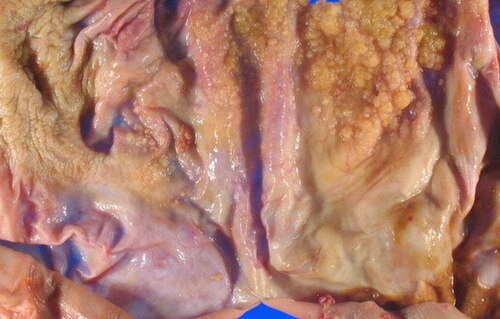
42 year old woman with a 9 cm ovarian mass
Site
ZZZ
Primary image
Low magnification. H&E stain.
Intermediate magnification
|
|
Intermediate magnification. H&E stain.
|
High magnification
|
|
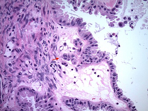
High magnification. H&E stain.
|
High magnification
|
|
High magnification. H&E stain.
|
Differential diagnosis
Differential diagnosis
|
|
SEROUS CYSTADENOFIBROMA OR ENDOCERVICAL TYPE BORDERLINE MUCINOUS TUMOR OR PAPILLARY CLEAR CELL CARCINOMA SIMULATING BORDERLINE TUMOR
|
Additional tests
More history
More history
|
|
MORE HISTORY HERE
|
Ask a colleague
Ask a colleague
|
|
CONSIDER DOING IMMUNO STAINS ?
|
Stains
| Alcian blue/PAS to Bilirubin |
|---|
| Test | Result |
|---|
| Alcian blue/PAS | Dr Torres would ask why! |
| Alican blue pH 1.0 | Dr Torres would ask why! |
| Alcian blue pH 2.5 | Dr Torres would ask why! |
| Auramine | Dr Torres would ask why! |
| Bielchowsky | Dr Torres would ask why! |
| Bilirubin | Dr Torres would ask why! |
|
|---|
|
| Colloidal iron to Fontana-Masson |
|---|
| Test | Result |
|---|
| Colloidal iron | Dr Torres would ask why! |
| Congo red | Dr Torres would ask why! |
| Cresyl violet | Dr Torres would ask why! |
| Dieterle | Dr Torres would ask why! |
| Diff Quik | Dr Torres would ask why! |
| Fontana-Masson | Dr Torres would ask why! |
|
|---|
|
| Gallyas to Gremelius |
|---|
| Test | Result |
|---|
| Gallyas | Dr Torres would ask why! |
| Giemsa | Dr Torres would ask why! |
| GMS | Dr Torres would ask why! |
| Gomori's trichrome | Dr Torres would ask why! |
| Gram | Dr Torres would ask why! |
| Gremelius | Dr Torres would ask why! |
|
|---|
|
| JMS to Mucicarmine |
|---|
| Test | Result |
|---|
| JMS | Dr Torres would ask why! |
| Jones | Dr Torres would ask why! |
| Kinyoun | Dr Torres would ask why! |
| Luxol fast blue | Dr Torres would ask why! |
| Masson trichrome | Dr Torres would ask why! |
| M-MAS | Dr Torres would ask why! |
| Movat | Dr Torres would ask why! |
| Mucicarmine | Dr Torres would ask why! |
|
|---|
|
| Oil red O to Prussian blue |
|---|
| Test | Result |
|---|
| Oil red O | Dr Torres would ask why! |
| Orecein | Dr Torres would ask why! |
| PAS | Dr Torres would ask why! |
| PASD | Dr Torres would ask why! |
| PASF | Dr Torres would ask why! |
| PTAH | Dr Torres would ask why! |
| Prussian blue | Dr Torres would ask why! |
|
|---|
|
| Reticulin to Ziehl-Neelsen |
|---|
| Test | Result |
|---|
| Reticulin | Dr Torres would ask why! |
| Sudan black B | Dr Torres would ask why! |
| Toluidine blue | Dr Torres would ask why! |
| Verhoeff | Dr Torres would ask why! |
| Von Kossa | Dr Torres would ask why! |
| Warthin-Starry | Dr Torres would ask why! |
| Ziehl-Neeslen | Dr Torres would ask why! |
|
|---|
|
IHC
| Alpha-1 AT to Cathepsin K |
|---|
| Test | Result |
|---|
| alpha-1 AT | Dr Torres would ask why! |
| ACTH | Dr Torres would ask why! |
| AE1/AE1 | Dr Torres would ask why! |
| alpha-fetoprotein | Dr Torres would ask why! |
| Alk-I | Dr Torres would ask why! |
| AMACR | Dr Torres would ask why! |
| AR | Dr Torres would ask why! |
| ATRX | Dr Torres would ask why! |
| Beta2-microglobulin | Dr Torres would ask why! |
| B72.3 | Dr Torres would ask why! |
| Beta-catenin | Dr Torres would ask why! |
| BCL2 | Dr Torres would ask why! |
| BCL6 | Dr Torres would ask why! |
| BCLxL | Dr Torres would ask why! |
| C3 comp | Dr Torres would ask why! |
| CA9 | Dr Torres would ask why! |
| Calcitonin | Dr Torres would ask why! |
| Calponin | Dr Torres would ask why! |
| Calretinin | Dr Torres would ask why! |
| CAM5.2 | Dr Torres would ask why! |
| Cathepsin K | Dr Torres would ask why! |
|
|---|
|
| CD10 to Chromogranin |
|---|
| Test | Result |
|---|
| CD10 | Dr Torres would ask why! |
| CD117 | Dr Torres would ask why! |
| CD138 | Dr Torres would ask why! |
| CD15 | Dr Torres would ask why! |
| CD1a | Dr Torres would ask why! |
| CD20 | Dr Torres would ask why! |
| CD21 | Dr Torres would ask why! |
| CD23 | Dr Torres would ask why! |
| CD3 | Dr Torres would ask why! |
| CD30 | Dr Torres would ask why! |
| CD31 | Dr Torres would ask why! |
| CD34 | Dr Torres would ask why! |
| CD35 | Dr Torres would ask why! |
| CD4 | Dr Torres would ask why! |
| CD43 | Dr Torres would ask why! |
| CD45 (LCA) | Dr Torres would ask why! |
| CD5 | Dr Torres would ask why! |
| CD56 | Dr Torres would ask why! |
| CD57 | Dr Torres would ask why! |
| CD68 | Dr Torres would ask why! |
| CD7 | Dr Torres would ask why! |
| CD79a | Dr Torres would ask why! |
| CD8 | Dr Torres would ask why! |
| CD99 | Dr Torres would ask why! |
| CDX2 | Dr Torres would ask why! |
| CEA-m | Dr Torres would ask why! |
| Chromogranin | Dr Torres would ask why! |
|
|---|
|
| CK17 to Glypican 3 |
|---|
| Test | Result |
|---|
| CK17 | Dr Torres would ask why! |
| CK19 | Dr Torres would ask why! |
| CK20 | Dr Torres would ask why! |
| CK34betaE12 | Dr Torres would ask why! |
| CK5/6 | Dr Torres would ask why! |
| CK7 | Dr Torres would ask why! |
| CMV | Dr Torres would ask why! |
| c-MYC | Dr Torres would ask why! |
| Cyclin D1 | Dr Torres would ask why! |
| D2-40 | Dr Torres would ask why! |
| Desmin | Dr Torres would ask why! |
| DOG1 | Dr Torres would ask why! |
| EBV | Dr Torres would ask why! |
| EMA | Dr Torres would ask why! |
| ER and PR | POSITIVE |
| Factor VIII | Dr Torres would ask why! |
| Factor XIIIa | Dr Torres would ask why! |
| Fascin | Dr Torres would ask why! |
| FH | Dr Torres would ask why! |
| FSH | Dr Torres would ask why! |
| Gastrin | Dr Torres would ask why! |
| GATA3 | Dr Torres would ask why! |
| GCDFP-15 (BRST2) | Dr Torres would ask why! |
| GFAP | Dr Torres would ask why! |
| GH | Dr Torres would ask why! |
| Glucagon | Dr Torres would ask why! |
| Glypican-3 | Dr Torres would ask why! |
|
|---|
|
| HBME-1 to IgM |
|---|
| Test | Result |
|---|
| HBME-1 | Dr Torres would ask why! |
| HBV core | Dr Torres would ask why! |
| HBV surface | Dr Torres would ask why! |
| H-caldesmon | Dr Torres would ask why! |
| HCG | Dr Torres would ask why! |
| Helicobacter | Dr Torres would ask why! |
| Hepatocyte | Dr Torres would ask why! |
| HER2/neu | Dr Torres would ask why! |
| HHV-8 | Dr Torres would ask why! |
| HMB-45 | Dr Torres would ask why! |
| HNF1beta | Dr Torres would ask why! |
| HPV | Dr Torres would ask why! |
| HSV-I | Dr Torres would ask why! |
| HSV-II | Dr Torres would ask why! |
| IDH-1 | Dr Torres would ask why! |
| Inhibin | Dr Torres would ask why! |
| INI1 (BAF47) | Dr Torres would ask why! |
| Insulin | Dr Torres would ask why! |
| Kappa | Dr Torres would ask why! |
| Ki-67 | Dr Torres would ask why! |
| Lambda | Dr Torres would ask why! |
| Leu 7 | Dr Torres would ask why! |
| IgA | Dr Torres would ask why! |
| IgG | Dr Torres would ask why! |
| IgM | Dr Torres would ask why! |
|
|---|
|
| LH to PDGFR |
|---|
| Test | Result |
|---|
| LH | Dr Torres would ask why! |
| LIN28 | Dr Torres would ask why! |
| Lysozyme | Dr Torres would ask why! |
| mammoglobin | Dr Torres would ask why! |
| MAP2 | Dr Torres would ask why! |
| MCV | Dr Torres would ask why! |
| Melanin A | Dr Torres would ask why! |
| MHC class I | Dr Torres would ask why! |
| MITF | Dr Torres would ask why! |
| MUM1 | Dr Torres would ask why! |
| Myeloperoxidase | Dr Torres would ask why! |
| MYO D1 | Dr Torres would ask why! |
| Myoglobin | Dr Torres would ask why! |
| Napsin | Dr Torres would ask why! |
| NF | Dr Torres would ask why! |
| NKX3.1 | Dr Torres would ask why! |
| NSE | Dr Torres would ask why! |
| OCT3/4 | Dr Torres would ask why! |
| p16 | Dr Torres would ask why! |
| P501S | Dr Torres would ask why! |
| p53 | Dr Torres would ask why! |
| p57 | Dr Torres would ask why! |
| p63 | Dr Torres would ask why! |
| Pankeratin | Dr Torres would ask why! |
| PAX2 | Dr Torres would ask why! |
| PAX5 | Dr Torres would ask why! |
| PAX8 | Dr Torres would ask why! |
| PCNA | Dr Torres would ask why! |
| PDGFR | Dr Torres would ask why! |
|
|---|
|
| PLAP to WT1 |
|---|
| Test | Result |
|---|
| PLAP | Dr Torres would ask why! |
| PNL-2C | Dr Torres would ask why! |
| Prolactin | Dr Torres would ask why! |
| PSA | Dr Torres would ask why! |
| PSAP | Dr Torres would ask why! |
| RCC | Dr Torres would ask why! |
| S-100 | Dr Torres would ask why! |
| SALL4 | Dr Torres would ask why! |
| Smooth muscle actin | Dr Torres would ask why! |
| Somatostatin | Dr Torres would ask why! |
| STAT6 | Dr Torres would ask why! |
| Synaptophysin | Dr Torres would ask why! |
| TdT | Dr Torres would ask why! |
| TFE3 | Dr Torres would ask why! |
| TFEB | Dr Torres would ask why! |
| Thyroglobulin | Dr Torres would ask why! |
| Toxoplasma | Dr Torres would ask why! |
| TSH | Dr Torres would ask why! |
| TTF-1 | Dr Torres would ask why! |
| Ubiquitin | Dr Torres would ask why! |
| UCHL1 (PGP9.5) | Dr Torres would ask why! |
| Ulex Europaeus | Dr Torres would ask why! |
| Vimentin | Dr Torres would ask why! |
| VIP | Dr Torres would ask why! |
| VZV | Dr Torres would ask why! |
| WT-1 | POSITIVE |
|
|---|
|
Molecular testing
Chromosomal translocations
| Translocations Chr 1-10 |
|---|
| Test | Result |
|---|
| t(1;13) PAX7-FKHR | Dr Torres would ask why! |
| t(2,13) PAX3-FKHR | Dr Torres would ask why! |
| t(8;14) MYC-IGH | Dr Torres would ask why! |
| t(9;22) BCR-ABL | Dr Torres would ask why! |
| t(9;22) CHN-EWS | Dr Torres would ask why! |
|
|---|
|
| Translocations Chr 11-13 |
|---|
| Test | Result |
|---|
| t(11;14) CCND1-IGH | Dr Torres would ask why! |
| t(11;22) EWS-WT1 | Dr Torres would ask why! |
| t(11;22) FLI1-EWS | Dr Torres would ask why! |
| t(12;15) ETV6-NTRK3 | Dr Torres would ask why! |
| t(12;16) FUS-ATF1 | Dr Torres would ask why! |
| t(12;16) CHOP-TLS | Dr Torres would ask why! |
| t(12;22) EWS-ATF1 | Dr Torres would ask why! |
|
|---|
|
| Translocations Chr 14-22 |
|---|
| Test | Result |
|---|
| t(14,18) IGH-BCL2 | Dr Torres would ask why! |
| t(15;17) PML-RARA | Dr Torres would ask why! |
| t(16;21) FUS-ERG | Dr Torres would ask why! |
| t(17;22) COLA1-PDGFB | Dr Torres would ask why! |
| t(21;22) EWS-ERG | Dr Torres would ask why! |
|
|---|
|
| Translocations Chr X & Y |
|---|
| Test | Result |
|---|
| t(X;1) PRCC-TFE3 | Dr Torres would ask why! |
| t(X;17) TFE3-ASPL | Dr Torres would ask why! |
| t(X;18) SYT-SSX | Dr Torres would ask why! |
|
|---|
|
Other molecular tests
| Molecular tests (A-B) |
|---|
| Test | Result |
|---|
| ALK sequencing | Dr Torres would ask why! |
| B cell clonality Southern / PCR | Dr Torres would ask why! |
| BCL2 PCR | Dr Torres would ask why! |
| BRAF sequencing | Dr Torres would ask why! |
|
|---|
|
| Molecular tests (C-H) |
|---|
| Test | Result |
|---|
| EBV PCR | Dr Torres would ask why! |
| EGRF sequencing | Dr Torres would ask why! |
| H3F3A sequencing | Dr Torres would ask why! |
| HHV-8 PCR | Dr Torres would ask why! |
|
|---|
|
| Molecular tests (I-J) |
|---|
| Test | Result |
|---|
| Identity testing PCR | Dr Torres would ask why! |
| IDH1/2 PCR | Dr Torres would ask why! |
| JAK2 V617F ARMS | Dr Torres would ask why! |
|
|---|
|
| Molecular tests (K-Z) |
|---|
| Test | Result |
|---|
| KIT sequencing | Dr Torres would ask why! |
| LOH 1p/19q PCR | Dr Torres would ask why! |
| T cell clonality Southern / PCR | Dr Torres would ask why! |
|
|---|
|
Diagnosis
Diagnosis
|
|
BORDERLINE SEROUS TUMOR OF THE OVARY WITH MICROINVASION.
The characteristic feature of borderline serous tumor is the hierarchical branching of the papillae, where the papillae progressively branch from larger ones to smaller one and finally into tufts of epithelial cells. The cells show only mild to moderate atypia. A cytadenofibroma may have similar proliferation, but by convention should be less than 5% of the tumor. Focal mucinous change may also occur. Papillary clear cell carcinoma simulating borderline tumor will show a WT1-ve, ER-ve, PR-ve immunoprofile in contrast to the positive profile of the serous tumor. Often clusters of cells with abundant eosinophilic cytoplasm are seen at the surface of papillae (star, image 2; oval image 3) or in lymphatic like spaces either as single cells or small papillary clusters. This theoretically represents microinvasion which is variably defined by different authors (<3 or 5 mm or 10mm2). However, recent evidence suggests that the two patterns of invasion have vastly different prognosis. The AEC (abundant eosinophilic cells) are p16 positive and reflect a terminally differentiated senescent phenotype. If invasion is exclusively composed of this cell type, then these do not portend a poor prognosis.[1]. These cells are significantly more often seen in BRAF mutated tumors, and are perhaps associated with a better prognosis. (Am J Surg Pathol 2014;38:1603–1611)
References
- ↑ Maniar, KP.; Wang, Y.; Visvanathan, K.; Shih, IeM.; Kurman, RJ. (Jun 2014). "Evaluation of microinvasion and lymph node involvement in ovarian serous borderline/atypical proliferative serous tumors: a morphologic and immunohistochemical analysis of 37 cases.". Am J Surg Pathol 38 (6): 743-55. doi:10.1097/PAS.0000000000000155. PMID 24441661.
|
Other cases
|
|---|
| | Number | |
|---|
| Subspecialty
(Difficulty) |
Autopsy pathology (jr,sr, f/e)
Breast pathology (jr,sr, f/e)
Cardiovascular pathology (jr,sr, f/e)
Cytopathology (jr,sr, f/e)
Dermatopathology (jr,sr, f/e)
Endocrine pathology (jr,sr, f/e)
Forensic pathology (jr,sr, f/e)
Gastrointestinal pathology (jr,sr, f/e)
Genitourinary pathology (jr,sr, f/e)
Gynecologic pathology (jr,sr, f/e)
Hematopathology (jr,sr, f/e)
Head and neck pathology (jr,sr, f/e)
Lymph node pathology (jr,sr, f/e)
Medical kidney pathology (jr,sr, f/e)
Molecular pathology (jr,sr, f/e)
Neuropathology (jr,sr, f/e)
Pediatric pathology (jr,sr, f/e)
Pulmonary pathology (jr,sr, f/e)
Placental pathology (jr,sr, f/e)
Soft tissue pathology (jr,sr, f/e) |
|---|
| | Difficulty | |
|---|
|


