Difference between revisions of "Sertoli-Leydig cell tumour"
Jump to navigation
Jump to search
(→IHC) |
|||
| Line 10: | Line 10: | ||
# Sertoli ''or'' Leydig cells.<ref name=Ref_PBoD1103>{{Ref PBoD|1103}}</ref> | # Sertoli ''or'' Leydig cells.<ref name=Ref_PBoD1103>{{Ref PBoD|1103}}</ref> | ||
#* Leydig cells: | #* Leydig cells: | ||
#** Abundant solid eosinophilic cytoplasm. | #**Polygonal pink cells | ||
#** Abundant solid or somewhat granular eosinophilic cytoplasm. | |||
#** Round nuclei with fine chromatin and a small or indistinct [[nucleolus]]. | #** Round nuclei with fine chromatin and a small or indistinct [[nucleolus]]. | ||
#** Often in small clusters ~ 5-25 cells/cluster. | #** Often in small clusters ~ 5-25 cells/cluster. | ||
| Line 19: | Line 20: | ||
# Stroma. | # Stroma. | ||
# +/- Sarcomatous features (mucinous glands, bone, cartilage). | # +/- Sarcomatous features (mucinous glands, bone, cartilage). | ||
*Well differentiated - | |||
**Mature Sertoli cells line form tubules that grow in a fibrous stroma containing clusters of Leydig | |||
cells | |||
*Intermediate to poorly differentiated - | |||
**A more disorganized, more cellular tumor with less mature Sertoli cells growing in trabeculae and nests. Some tubule formation, either round or retiform. Leydig cells, either singly or in clusters, are present in a cellular stroma. | |||
DDx: | DDx: | ||
Revision as of 10:33, 21 March 2015
Sertoli-Leydig cell tumour, also Sertoli-Leydig tumour, is a rare tumour of the gonad in the sex cord-stromal group of tumours.
General
- Sertoli and leydig cells are normal in the testis.
- Poorly differentiated tumours have sarcomatous features.[1]
- May present with masculinization (virilization).[2]
Microscopic
Features:
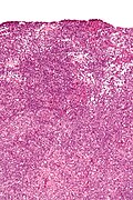
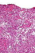
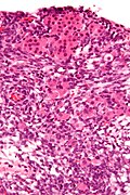
- Sertoli or Leydig cells.[1]
- Leydig cells:
- Polygonal pink cells
- Abundant solid or somewhat granular eosinophilic cytoplasm.
- Round nuclei with fine chromatin and a small or indistinct nucleolus.
- Often in small clusters ~ 5-25 cells/cluster.
- Sertoli cells:
- Pale/clear vacuolated cytoplasm.
- Irregular nuclei with irregular/vacuolated-appearing chromatin.
- Architecture: tubules, cords or sheets.
- Leydig cells:
- Stroma.
- +/- Sarcomatous features (mucinous glands, bone, cartilage).
- Well differentiated -
- Mature Sertoli cells line form tubules that grow in a fibrous stroma containing clusters of Leydig
cells
- Intermediate to poorly differentiated -
- A more disorganized, more cellular tumor with less mature Sertoli cells growing in trabeculae and nests. Some tubule formation, either round or retiform. Leydig cells, either singly or in clusters, are present in a cellular stroma.
DDx:
- Endometrioid carcinoma of the ovary.
- Luteinized adult granulosa cell tumour - super rare, 50% of cell with eosinophilic cytoplasm, other findings of granulosa cell tumour, e.g. Call-Exner bodies.[3]
Images
www:
IHC
Features:[4]
- AE1/AE3 +ve
- Inhibin +ve
- WT-1 +ve.
- Melan A (MART-1) +ve - marks the Leydig component.
- Vimentin +ve.[5]
- Calretinin +ve.
- CD99 +ve.
Others:[5]
- CD34 -ve.
- EMA -ve.
Pan-keratins and AE1/AE3 may mark granulosa cell tumors and Sertoli cell tumors causing confusion with adenocarcinoma. EMA is a better marker to exclude an epithelial tumor as EMA is negative in sex cord-stromal tumors. Adding complexity, endometrioid adenocarcinomas may occasionally weakly express inhibin, calretinin or WT-1.
See also
References
- ↑ 1.0 1.1 Cotran, Ramzi S.; Kumar, Vinay; Fausto, Nelson; Nelso Fausto; Robbins, Stanley L.; Abbas, Abul K. (2005). Robbins and Cotran pathologic basis of disease (7th ed.). St. Louis, Mo: Elsevier Saunders. pp. 1103. ISBN 0-7216-0187-1.
- ↑ Xiao, H.; Li, B.; Zuo, J.; Feng, X.; Li, X.; Zhang, R.; Wu, L. (Mar 2013). "Ovarian Sertoli-Leydig cell tumor: a report of seven cases and a review of the literature.". Gynecol Endocrinol 29 (3): 192-5. doi:10.3109/09513590.2012.738723. PMID 23173550.
- ↑ Ganesan, R.; Hirschowitz, L.; Baltrušaitytė, I.; McCluggage, WG. (Sep 2011). "Luteinized adult granulosa cell tumor--a series of 9 cases: revisiting a rare variant of adult granulosa cell tumor.". Int J Gynecol Pathol 30 (5): 452-9. doi:10.1097/PGP.0b013e318214b17f. PMID 21804396.
- ↑ Zhao, C.; Vinh, TN.; McManus, K.; Dabbs, D.; Barner, R.; Vang, R. (Mar 2009). "Identification of the most sensitive and robust immunohistochemical markers in different categories of ovarian sex cord-stromal tumors.". Am J Surg Pathol 33 (3): 354-66. doi:10.1097/PAS.0b013e318188373d. PMID 19033865.
- ↑ 5.0 5.1 Kondi-Pafiti, A.; Grapsa, D.; Kairi-Vassilatou, E.; Carvounis, E.; Hasiakos, D.; Kontogianni, K.; Fotiou, S. (2010). "Granulosa cell tumors of the ovary: a clinicopathologic and immunohistochemical study of 21 cases.". Eur J Gynaecol Oncol 31 (1): 94-8. PMID 20349790.


