Difference between revisions of "Placental site nodule"
Jump to navigation
Jump to search
| Line 71: | Line 71: | ||
{{Reflist|1}} | {{Reflist|1}} | ||
[[Category:Gestational trophoblastic disease]] | |||
[[Category:Diagnosis]] | [[Category:Diagnosis]] | ||
Revision as of 15:46, 22 May 2014
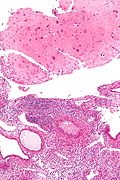
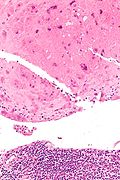
Placental site nodule, abbreviated PSN, is a benign remnant of a placenta. It is seen on endometrial biopsies and hysterectomy specimens.
Implantation site redirects here.
General
- Benign.
- Intermediate trophoblast remnants from a previous gestation.[1]
- Usually an incidental finding.
Clinical:
- Usually asymptomatic.
- Vaginal bleeding. (?)
- Infertility. (?)
Microscopic
Features:[1]
- Paucicellular with hyaline material scattered cells.
- Variable cell population:
- Small-large cells.
- Clear to eosinophilic cytoplasm.
- +/-Multinucleation.
Notes:
- No mitotic activity.
DDx:
- Invasive (cervical) squamous cell carcinoma.
- Can be sorted-out with IHC (SCC will typically be: p16 +ve, MIB1 +ve).
- Exaggerated placental site.
- Different histomorphology than PSN; EPS:[1] syncytiotrophoblastic tissue, in cords/nests, no hyaline nodules.
- Fibrin +/-inflammatory cells.
Images
www:
- PSN (ijpmonline.org).[1]
- PSN (gfmer.ch) - includes images from Jacob and Mohapatra.[1]
IHC
Features:[1]
- Inhibin alpha +ve.
- CK18 +ve.
- MIB1 low.
Other:
- p16 -ve.
Sign out
- CONSISTENT WITH MENSTRUAL ENDOMETRIUM (FRAGMENTED ENDOMETRIUM WITH SIMPLE
  GLANDS WITH APOPTOTIC CELLS, ABUNDANT NEUTROPHILS, CONDENSED ENDOMETRIAL STROMA
  (FOCAL) AND BLOOD).
- PLACENTAL SITE NODULE AND DECIDUA.
- NEGATIVE FOR HYPERPLASIA AND NEGATIVE FOR MALIGNANCY.
ENDOMETRIUM, BIOPSY:
- PROLIFERATIVE PHASE ENDOMETRIUM.
- BENIGN PLACENTAL SITE NODULE, SMALL.
- NEGATIVE FOR HYPERPLASIA.
See also
References
- ↑ 1.0 1.1 1.2 1.3 1.4 1.5 Jacob, S.; Mohapatra, D.. "Placental site nodule: a tumor-like trophoblastic lesion.". Indian J Pathol Microbiol 52 (2): 240-1. PMID 19332926. http://www.ijpmonline.org/text.asp?2009/52/2/240/48931.

