Difference between revisions of "Gallbladder carcinoma"
Jump to navigation
Jump to search
m (→Images: touch) |
(touch) |
||
| Line 1: | Line 1: | ||
{{ Infobox diagnosis | {{ Infobox diagnosis | ||
| Name = {{PAGENAME}} | | Name = {{PAGENAME}} | ||
| Image = Gallbladder adenocarcinoma (2) histopathology.jpg | | Image = Gallbladder adenocarcinoma (2) histopathology.jpg | ||
| Width = | | Width = | ||
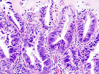
| Caption = Gallbladder adenocarcinoma. [[H&E stain]]. | | Caption = Gallbladder adenocarcinoma. [[H&E stain]]. | ||
Revision as of 05:12, 11 June 2015
| Gallbladder carcinoma | |
|---|---|
| Diagnosis in short | |
 Gallbladder adenocarcinoma. H&E stain. | |
|
| |
| LM | atypical epithelium usually gland forming, may be bland with an infiltrative growth pattern |
| LM DDx | adenomyoma of the gallbladder, metastatic carcinoma (e.g. cholangiocarcinoma), gallbladder adenoma |
| IHC | CK7 +ve, CK20 -ve, CDX2 -ve |
| Gross | lesion - usually fundus of gallbladder |
| Site | gallbladder |
|
| |
| Associated Dx | chronic cholecystitis, gallstones, primary sclerosing cholangitis, intestinal metaplasia of the gallbladder |
| Prevalence | uncommon |
Gallbladder carcinoma is a malignant epithelial neoplasm arising from the gallbladder. Most gallbladder carcinomas are adenocarcinomas.
General
- Uncommon.
Treatment:
- Cholecystectomy +/- lymph nodes +/- partial hepatectomy.[1]
Epidemiology
- Associated with gallstones.
- Increased risk in primary sclerosing cholangitis.
- Sex: female > male.
- Location: usually fundus, sometimes body.
Notes:
- Diffuse calcification of gallbladder wall, AKA "porcelain gallbladder" is not associated with carcinoma - based on a series of 10,741 cholecystectomies.[2]
- Focal mucosal calcification is associated with malignancy.[3]
- Cholangiocarcinoma is dealt with in the liver neoplasms article.
Gross
- Classic: mass projecting into the lumen.
- Marked gallbladder wall thickening.
- >10 mm should be considered with suspicion.[4]
Image:
Microscopic
Features:
- Usually adenocarcinoma.
- Mimics appearance of pancreatic ductal adenocarcinoma -- but less cellular mucin.[5]
Notes:
- May be very subtle, i.e. difficult to differentiate from normal glands.
- "Deep glands" that look bland shouldn't immediately be dismissed as benign.
Subtypes:
DDx:
- Adenomyoma of the gallbladder.
- Gallbladder adenoma.
- Metastatic carcinoma.
- Chronic cholecystitis with glands extending deep into the wall.
Images
www:
IHC
Features - conventional:[6]
- CK7 +ve (7 of 8 cases).
- CK20 -ve (7 of 8 cases).
- CDX2 -ve (8 of 8 cases).
See also
References
- ↑ Biswas, PK. (Jul 2010). "Carcinoma gallbladder.". Mymensingh Med J 19 (3): 477-81. PMID 20639849.
- ↑ Towfigh S, McFadden DW, Cortina GR, et al (January 2001). "Porcelain gallbladder is not associated with gallbladder carcinoma". Am Surg 67 (1): 7?0. PMID 11206901.
- ↑ Stephen, AE.; Berger, DL. (Jun 2001). "Carcinoma in the porcelain gallbladder: a relationship revisited.". Surgery 129 (6): 699-703. doi:10.1067/msy.2001.113888. PMID 11391368.
- ↑ Kim, HJ.; Park, JH.; Park, DI.; Cho, YK.; Sohn, CI.; Jeon, WK.; Kim, BI.; Choi, SH. (Feb 2012). "Clinical usefulness of endoscopic ultrasonography in the differential diagnosis of gallbladder wall thickening.". Dig Dis Sci 57 (2): 508-15. doi:10.1007/s10620-011-1870-0. PMID 21879282.
- ↑ Tadrous, Paul.J. Diagnostic Criteria Handbook in Histopathology: A Surgical Pathology Vade Mecum (1st ed.). Wiley. pp. 174. ISBN 978-0470519035.
- ↑ 6.0 6.1 Dursun, N.; Escalona, OT.; Roa, JC.; Basturk, O.; Bagci, P.; Cakir, A.; Cheng, J.; Sarmiento, J. et al. (Nov 2012). "Mucinous carcinomas of the gallbladder: clinicopathologic analysis of 15 cases identified in 606 carcinomas.". Arch Pathol Lab Med 136 (11): 1347-58. doi:10.5858/arpa.2011-0447-OA. PMID 23106580.
- ↑ Giang, TH.; Ngoc, TT.; Hassell, LA. (2012). "Carcinoma involving the gallbladder: a retrospective review of 23 cases - pitfalls in diagnosis of gallbladder carcinoma.". Diagn Pathol 7: 10. doi:10.1186/1746-1596-7-10. PMID 22284391.


