Difference between revisions of "Inflammatory fibroid polyp"
Jump to navigation
Jump to search
| Line 20: | Line 20: | ||
| Signs = | | Signs = | ||
| Symptoms = | | Symptoms = | ||
| Prevalence = | | Prevalence = uncommon | ||
| Bloodwork = | | Bloodwork = | ||
| Rads = | | Rads = | ||
| Line 34: | Line 34: | ||
*Through-out GI tract. | *Through-out GI tract. | ||
*Can be thought of as granulation tissue-like.<ref name=Ref_DCHH138/> | *Can be thought of as granulation tissue-like.<ref name=Ref_DCHH138/> | ||
*Uncommon. | |||
==Microscopic== | ==Microscopic== | ||
Revision as of 16:00, 17 November 2014
| Inflammatory fibroid polyp | |
|---|---|
| Diagnosis in short | |
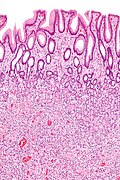
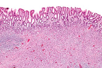 Inflammatory fibroid polyp. H&E stain. | |
|
| |
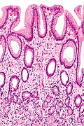
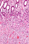
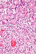
| LM | proliferating spindle cells - loosely arranged round blood vessels with perivascular hypocellular zones, eosinophils (usually prominent), +/-leiomyoma/schwannoma-like areas - with nuclear palisading |
| LM DDx | inflammatory myofibroblastic tumour, GIST, hemangiopericytoma, eosinophilic gastritis, schwannoma |
| IHC | CD34 +ve, vimentin +ve, CD117 -ve, S-100 -ve |
| Molecular | +/-PDGFRA mutations |
| Site | stomach, other places in the GI tract |
|
| |
| Prevalence | uncommon |
| Prognosis | good - benign |
Inflammatory fibroid polyp, abbreviated IFP, is an uncommon gastrointestinal polyp.
General
- Benign.
- Through-out GI tract.
- Can be thought of as granulation tissue-like.[1]
- Uncommon.
Microscopic
Features:[2]
- Proliferating spindle cells (fibroid) - key feature.
- Inflammation:
- Eosinophils - often prominent.
- +/-Leiomyoma/schwannoma-like areas - with nuclear palisading.[1]
- +/-Vascular for fibrous tissue.
- Poorly circumscribed/infiltrates into the lamina propria.
DDx:[4]
- Inflammatory myofibroblastic tumour.
- GIST - usually sharply demarcated border.
- Hemangiopericytoma.
- Eosinophilic gastritis.
Notes:
- Concentric = share the same centre.[5]
Images
IHC
Features:[2]
- CD34 +ve.
- There is a CD34 -ve variant.
- Vimentin +ve -- diffuse.[6]
Others:
- CD117 -ve.[7]
- S100 -ve.
Molecular
- A subset have mutations in PDGFRA.[2]
See also
References
- ↑ 1.0 1.1 1.2 Tadrous, Paul.J. Diagnostic Criteria Handbook in Histopathology: A Surgical Pathology Vade Mecum (1st ed.). Wiley. pp. 138. ISBN 978-0470519035.
- ↑ 2.0 2.1 2.2 Daum, O.; Hatlova, J.; Mandys, V.; Grossmann, P.; Mukensnabl, P.; Benes, Z.; Michal, M. (May 2010). "Comparison of morphological, immunohistochemical, and molecular genetic features of inflammatory fibroid polyps (Vanek's tumors).". Virchows Arch 456 (5): 491-7. doi:10.1007/s00428-010-0914-8. PMID 20393746.
- ↑ Iacobuzio-Donahue, Christine A.; Montgomery, Elizabeth A. (2005). Gastrointestinal and Liver Pathology: A Volume in the Foundations in Diagnostic Pathology Series (1st ed.). Churchill Livingstone. pp. 115. ISBN 978-0443066573.
- ↑ Rossi, P.; Montuori, M.; Balassone, V.; Ricciardi, E.; Anemona, L.; Manzelli, A.; Petrella, G.. "Inflammatory fibroid polyp. A case report and review of the literature.". Ann Ital Chir 83 (4): 347-51. PMID 22759473.
- ↑ URL: http://dictionary.reference.com/browse/concentric. Accessed on: 29 November 2011.
- ↑ Kolodziejczyk, P.; Yao, T.; Tsuneyoshi, M. (Nov 1993). "Inflammatory fibroid polyp of the stomach. A special reference to an immunohistochemical profile of 42 cases.". Am J Surg Pathol 17 (11): 1159-68. PMID 8214261.
- ↑ Ozolek, JA.; Sasatomi, E.; Swalsky, PA.; Rao, U.; Krasinskas, A.; Finkelstein, SD. (Mar 2004). "Inflammatory fibroid polyps of the gastrointestinal tract: clinical, pathologic, and molecular characteristics.". Appl Immunohistochem Mol Morphol 12 (1): 59-66. PMID 15163021.

