Difference between revisions of "Solitary fibrous tumour"
Jump to navigation
Jump to search
m (chg redirect) |
Jensflorian (talk | contribs) (→Images: STAT6 added.) |
||
| (13 intermediate revisions by 3 users not shown) | |||
| Line 1: | Line 1: | ||
{{ Infobox diagnosis | |||
| Name = {{PAGENAME}} | |||
| Image = Solitary_fibrous_tumour_high_mag.jpg | |||
| Width = | |||
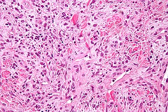
| Caption = Solitary fibrous tumour. [[H&E stain]]. | |||
| Micro = spindle cells in a patternless pattern, hemangiopericytoma-like areas ([[staghorn vessels]]), keloid-like collagen bundles, +/-well-circumscribed (common) | |||
| Subtypes = benign (common), malignant (uncommon) | |||
| LMDDx = | |||
| Stains = | |||
| IHC = CD34 ~90% +ve, CD99 ~70% +ve, BCL2 ~50% +ve | |||
| EM = | |||
| Molecular = | |||
| IF = | |||
| Gross = | |||
| Grossing = | |||
| Site = [[soft tissue lesions|soft tissue]] - [[fibroblastic/myofibroblastic tumours]], pleura | |||
| Assdx = | |||
| Syndromes = Doege-Potter syndrome | |||
| Clinicalhx = | |||
| Signs = | |||
| Symptoms = | |||
| Prevalence = | |||
| Bloodwork = | |||
| Rads = | |||
| Endoscopy = | |||
| Prognosis = usu. good | |||
| Other = | |||
| ClinDDx = | |||
}} | |||
'''Solitary fibrous tumour''', abbreviated '''SFT''', is a type of [[soft tissue lesion|soft tissue tumour]] that fits in the [[fibroblastic/myofibroblastic tumours]]. It is usually benign. | |||
SFT of the pleura is dealt with in a separate article ''[[solitary fibrous tumour of the pleura]]''. | |||
==General== | |||
*Grouped with ''hemangiopericytoma'' in the WHO classification - as it is thought to be the same tumour because both share the same molecular alteration.<ref name=Ref_WMSP609>{{Ref WMSP|609}}</ref><ref>{{Cite journal | last1 = Schweizer | first1 = L. | last2 = Koelsche | first2 = C. | last3 = Sahm | first3 = F. | last4 = Piro | first4 = RM. | last5 = Capper | first5 = D. | last6 = Reuss | first6 = DE. | last7 = Pusch | first7 = S. | last8 = Habel | first8 = A. | last9 = Meyer | first9 = J. | title = Meningeal hemangiopericytoma and solitary fibrous tumors carry the NAB2-STAT6 fusion and can be diagnosed by nuclear expression of STAT6 protein. | journal = Acta Neuropathol | volume = 125 | issue = 5 | pages = 651-8 | month = May | year = 2013 | doi = 10.1007/s00401-013-1117-6 | PMID = 23575898 }}</ref> | |||
*May be benign ''or'' malignant; more commonly benign.<ref>URL: [http://www.pathconsultddx.com/pathCon/diagnosis?pii=S1559-8675%2806%2970528-9 http://www.pathconsultddx.com/pathCon/diagnosis?pii=S1559-8675%2806%2970528-9]. Accessed on: 25 June 2010.</ref><ref>URL: [http://wjso.com/content/6/1/86 http://wjso.com/content/6/1/86]. Accessed on: 25 June 2010.</ref> | |||
*May be associated with hypoglycemia. | |||
**Known as ''Doege-Potter syndrome''.<ref name=pmid1474302>{{Cite journal | last1 = Roy | first1 = TM. | last2 = Burns | first2 = MV. | last3 = Overly | first3 = DJ. | last4 = Curd | first4 = BT. | title = Solitary fibrous tumor of the pleura with hypoglycemia: the Doege-Potter syndrome. | journal = J Ky Med Assoc | volume = 90 | issue = 11 | pages = 557-60 | month = Nov | year = 1992 | doi = | PMID = 1474302 }}</ref> | |||
*Leptomeningeal SFTs/hemangiopericytomas are classified as follows: | |||
** WHO grade I: classical SFT | |||
** WHO grade II: classical [[hemangiopericytoma]] | |||
** WHO grade III: anaplastic hemangiopericytoma / malignant SFT | |||
==Gross== | |||
*Soft tissue mass. | |||
==Microscopic== | |||
Features - benign: | |||
*Spindle cells in a patternless pattern. | |||
**Occasionally epithelioid cells - rare.<ref name=pmid17577399>{{Cite journal | last1 = Martorell | first1 = M. | last2 = Pérez-Vallés | first2 = A. | last3 = Gozalbo | first3 = F. | last4 = Garcia-Garcia | first4 = JA. | last5 = Gutierrez | first5 = J. | last6 = Gaona | first6 = J. | title = Solitary fibrous tumor of the thigh with epithelioid features: a case report. | journal = Diagn Pathol | volume = 2 | issue = | pages = 19 | month = | year = 2007 | doi = 10.1186/1746-1596-2-19 | PMID = 17577399 }}</ref> | |||
*Hemangiopericytoma-like area ([[staghorn vessels]]). | |||
*Keloid-like collagen bundles - '''key feature'''. | |||
*+/-Well-circumscribed (common). | |||
Criteria for malignancy:<ref name=Ref_WMSP609>{{Ref WMSP|609}}</ref> | |||
*Necrosis. | |||
*Mitoses >4/10 HPF -- definition suffers from [[HPFitis]]. | |||
*Increased cellularity. | |||
*Marked nuclear atypia. | |||
*Infiltrative margin. | |||
===Images=== | |||
<gallery> | |||
Image:Solitary_fibrous_tumour_low_mag.jpg | Benign SFT - low mag. (WC) | |||
Image:Solitary_fibrous_tumour_intermed_mag.jpg | Benign SFT - intermed. mag. (WC) | |||
Image:Solitary_fibrous_tumour_high_mag.jpg | Benign SFT - high mag. (WC) | |||
File:Neuropathology_case_VI_01.jpg | Nuclear STAT6 immunoreactivity in malignant SFT (WC/jensflorian) | |||
</gallery> | |||
www: | |||
*[http://path.upmc.edu/cases/case272.html SFT of the brain - several images (upmc.edu)]. | |||
Malignant solitary fibrous tumor (low to intermediate grade tumor). 15 cm mass in upper arm of 50 year old man.<ref>Am J Surg Pathol 2014;38:552-559</ref> | |||
<gallery> | |||
File:3 18039322534203 sl 1.png|malignant solitary fibrous tumor | Hypocellular (green arrow) and hypercellular (cyan arrow) areas alternate. | |||
File:3 18039322534203 sl 2.png|malignant solitary fibrous tumor | Vascular spaces suggest antlers (staghorn). Note subcapsular hemorrhage. | |||
File:3 18039322534203 sl 3.png|malignant solitary fibrous tumor | Stromal hyalinization | |||
File:3 18039322534203 sl 4.png|malignant solitary fibrous tumor | Nuclei, lacking organization, show varied size and shape, with some cells being round to ovoid. The arrow points to myxoid focus in stroma. When a majority of the following are present, a low to intermediate grade malignant solitary fibrous tumor is present: 1) size > 5 cm (this case), 2) pleomorphism with round cells/epithelioid cells (this case), 3) subcapsular hemorrhage (this case), 4) ≥4 mitoses per 10 high power fields, 5) necrosis with perivascular tumor sparing. STAT 6 positivity is very helpful in separating this tumor from its mimics | |||
</gallery> | |||
==IHC== | |||
*CD34 ~90% +ve. | |||
*CD99 ~70% +ve. | |||
*BCL2 ~50% +ve. | |||
*Stat6 nuclear +ve.<ref>{{Cite journal | last1 = Cheah | first1 = AL. | last2 = Billings | first2 = SD. | last3 = Goldblum | first3 = JR. | last4 = Carver | first4 = P. | last5 = Tanas | first5 = MZ. | last6 = Rubin | first6 = BP. | title = STAT6 rabbit monoclonal antibody is a robust diagnostic tool for the distinction of solitary fibrous tumour from its mimics. | journal = Pathology | volume = 46 | issue = 5 | pages = 389-95 | month = Aug | year = 2014 | doi = 10.1097/PAT.0000000000000122 | PMID = 24977739 }}</ref> | |||
==Molecular== | |||
*NAB2/STAT6 fusions.<ref>{{Cite journal | last1 = Mohajeri | first1 = A. | last2 = Tayebwa | first2 = J. | last3 = Collin | first3 = A. | last4 = Nilsson | first4 = J. | last5 = Magnusson | first5 = L. | last6 = von Steyern | first6 = FV. | last7 = Brosjö | first7 = O. | last8 = Domanski | first8 = HA. | last9 = Larsson | first9 = O. | title = Comprehensive genetic analysis identifies a pathognomonic NAB2/STAT6 fusion gene, nonrandom secondary genomic imbalances, and a characteristic gene expression profile in solitary fibrous tumor. | journal = Genes Chromosomes Cancer | volume = 52 | issue = 10 | pages = 873-86 | month = Oct | year = 2013 | doi = 10.1002/gcc.22083 | PMID = 23761323 }}</ref><ref>{{Cite journal | last1 = Robinson | first1 = DR. | last2 = Wu | first2 = YM. | last3 = Kalyana-Sundaram | first3 = S. | last4 = Cao | first4 = X. | last5 = Lonigro | first5 = RJ. | last6 = Sung | first6 = YS. | last7 = Chen | first7 = CL. | last8 = Zhang | first8 = L. | last9 = Wang | first9 = R. | title = Identification of recurrent NAB2-STAT6 gene fusions in solitary fibrous tumor by integrative sequencing. | journal = Nat Genet | volume = 45 | issue = 2 | pages = 180-5 | month = Feb | year = 2013 | doi = 10.1038/ng.2509 | PMID = 23313952 }}</ref> | |||
==DDx== | |||
*[[Meningioma]] | |||
*Cellular [[angiofibroma]] | |||
*[[Myofibroblastoma]] | |||
*Benign [[fibrous histiocytoma]] | |||
*[[Dermatofibrosarcoma protruberans]] | |||
*[[Fibromyxoid sarcoma]] | |||
==See also== | |||
*[[Fibroblastic/myofibroblastic tumours]]. | |||
*[[Hemangiopericytoma]] | |||
==References== | |||
{{Reflist|2}} | |||
[[Category:Fibroblastic/myofibroblastic tumours]] | |||
[[Category:Diagnosis]] | |||
[[Category:Neuropathology]] | |||
Latest revision as of 07:21, 15 December 2016
| Solitary fibrous tumour | |
|---|---|
| Diagnosis in short | |
 Solitary fibrous tumour. H&E stain. | |
|
| |
| LM | spindle cells in a patternless pattern, hemangiopericytoma-like areas (staghorn vessels), keloid-like collagen bundles, +/-well-circumscribed (common) |
| Subtypes | benign (common), malignant (uncommon) |
| IHC | CD34 ~90% +ve, CD99 ~70% +ve, BCL2 ~50% +ve |
| Site | soft tissue - fibroblastic/myofibroblastic tumours, pleura |
|
| |
| Syndromes | Doege-Potter syndrome |
|
| |
| Prognosis | usu. good |
Solitary fibrous tumour, abbreviated SFT, is a type of soft tissue tumour that fits in the fibroblastic/myofibroblastic tumours. It is usually benign.
SFT of the pleura is dealt with in a separate article solitary fibrous tumour of the pleura.
General
- Grouped with hemangiopericytoma in the WHO classification - as it is thought to be the same tumour because both share the same molecular alteration.[1][2]
- May be benign or malignant; more commonly benign.[3][4]
- May be associated with hypoglycemia.
- Known as Doege-Potter syndrome.[5]
- Leptomeningeal SFTs/hemangiopericytomas are classified as follows:
- WHO grade I: classical SFT
- WHO grade II: classical hemangiopericytoma
- WHO grade III: anaplastic hemangiopericytoma / malignant SFT
Gross
- Soft tissue mass.
Microscopic
Features - benign:
- Spindle cells in a patternless pattern.
- Occasionally epithelioid cells - rare.[6]
- Hemangiopericytoma-like area (staghorn vessels).
- Keloid-like collagen bundles - key feature.
- +/-Well-circumscribed (common).
Criteria for malignancy:[1]
- Necrosis.
- Mitoses >4/10 HPF -- definition suffers from HPFitis.
- Increased cellularity.
- Marked nuclear atypia.
- Infiltrative margin.
Images
www:
Malignant solitary fibrous tumor (low to intermediate grade tumor). 15 cm mass in upper arm of 50 year old man.[7]
Nuclei, lacking organization, show varied size and shape, with some cells being round to ovoid. The arrow points to myxoid focus in stroma. When a majority of the following are present, a low to intermediate grade malignant solitary fibrous tumor is present: 1) size > 5 cm (this case), 2) pleomorphism with round cells/epithelioid cells (this case), 3) subcapsular hemorrhage (this case), 4) ≥4 mitoses per 10 high power fields, 5) necrosis with perivascular tumor sparing. STAT 6 positivity is very helpful in separating this tumor from its mimics
IHC
- CD34 ~90% +ve.
- CD99 ~70% +ve.
- BCL2 ~50% +ve.
- Stat6 nuclear +ve.[8]
Molecular
DDx
- Meningioma
- Cellular angiofibroma
- Myofibroblastoma
- Benign fibrous histiocytoma
- Dermatofibrosarcoma protruberans
- Fibromyxoid sarcoma
See also
References
- ↑ 1.0 1.1 Humphrey, Peter A; Dehner, Louis P; Pfeifer, John D (2008). The Washington Manual of Surgical Pathology (1st ed.). Lippincott Williams & Wilkins. pp. 609. ISBN 978-0781765275.
- ↑ Schweizer, L.; Koelsche, C.; Sahm, F.; Piro, RM.; Capper, D.; Reuss, DE.; Pusch, S.; Habel, A. et al. (May 2013). "Meningeal hemangiopericytoma and solitary fibrous tumors carry the NAB2-STAT6 fusion and can be diagnosed by nuclear expression of STAT6 protein.". Acta Neuropathol 125 (5): 651-8. doi:10.1007/s00401-013-1117-6. PMID 23575898.
- ↑ URL: http://www.pathconsultddx.com/pathCon/diagnosis?pii=S1559-8675%2806%2970528-9. Accessed on: 25 June 2010.
- ↑ URL: http://wjso.com/content/6/1/86. Accessed on: 25 June 2010.
- ↑ Roy, TM.; Burns, MV.; Overly, DJ.; Curd, BT. (Nov 1992). "Solitary fibrous tumor of the pleura with hypoglycemia: the Doege-Potter syndrome.". J Ky Med Assoc 90 (11): 557-60. PMID 1474302.
- ↑ Martorell, M.; Pérez-Vallés, A.; Gozalbo, F.; Garcia-Garcia, JA.; Gutierrez, J.; Gaona, J. (2007). "Solitary fibrous tumor of the thigh with epithelioid features: a case report.". Diagn Pathol 2: 19. doi:10.1186/1746-1596-2-19. PMID 17577399.
- ↑ Am J Surg Pathol 2014;38:552-559
- ↑ Cheah, AL.; Billings, SD.; Goldblum, JR.; Carver, P.; Tanas, MZ.; Rubin, BP. (Aug 2014). "STAT6 rabbit monoclonal antibody is a robust diagnostic tool for the distinction of solitary fibrous tumour from its mimics.". Pathology 46 (5): 389-95. doi:10.1097/PAT.0000000000000122. PMID 24977739.
- ↑ Mohajeri, A.; Tayebwa, J.; Collin, A.; Nilsson, J.; Magnusson, L.; von Steyern, FV.; Brosjö, O.; Domanski, HA. et al. (Oct 2013). "Comprehensive genetic analysis identifies a pathognomonic NAB2/STAT6 fusion gene, nonrandom secondary genomic imbalances, and a characteristic gene expression profile in solitary fibrous tumor.". Genes Chromosomes Cancer 52 (10): 873-86. doi:10.1002/gcc.22083. PMID 23761323.
- ↑ Robinson, DR.; Wu, YM.; Kalyana-Sundaram, S.; Cao, X.; Lonigro, RJ.; Sung, YS.; Chen, CL.; Zhang, L. et al. (Feb 2013). "Identification of recurrent NAB2-STAT6 gene fusions in solitary fibrous tumor by integrative sequencing.". Nat Genet 45 (2): 180-5. doi:10.1038/ng.2509. PMID 23313952.


