Difference between revisions of "P53"
Jump to navigation
Jump to search
Jensflorian (talk | contribs) (→Interpretation: +neuropathology) |
|||
| (2 intermediate revisions by one other user not shown) | |||
| Line 11: | Line 11: | ||
| Subspecial = | | Subspecial = | ||
| Pattern = | | Pattern = | ||
| Positive = nuclear staining | | Positive = nuclear staining (>60% or completely negative - see ''interpretation'') | ||
| Negative = | | Negative = | ||
| Other = | | Other = | ||
}} | }} | ||
'''p53''' | '''p53''' marks a [[tumour suppressor]] protein commonly implicated in [[cancer]] and a common [[immunostain]]. | ||
==Interpretation== | ==Interpretation== | ||
*p53 may be one of the most misinterpreted stains. | *p53 may be one of the most misinterpreted stains. | ||
**TP53 mutations are associated with >60% staining and no staining (0% of cell labelled).<ref name=pmid21552211 >{{Cite journal | last1 = Yemelyanova | first1 = A. | last2 = Vang | first2 = R. | last3 = Kshirsagar | first3 = M. | last4 = Lu | first4 = D. | last5 = Marks | first5 = MA. | last6 = Shih | first6 = IeM. | last7 = Kurman | first7 = RJ. | title = Immunohistochemical staining patterns of p53 can serve as a surrogate marker for TP53 mutations in ovarian carcinoma: an immunohistochemical and nucleotide sequencing analysis. | journal = Mod Pathol | volume = 24 | issue = 9 | pages = 1248-53 | month = Sep | year = 2011 | doi = 10.1038/modpathol.2011.85 | PMID = 21552211 }}</ref> | **TP53 mutations are associated with >60% staining and no staining (0% of cell labelled).<ref name=pmid21552211 >{{Cite journal | last1 = Yemelyanova | first1 = A. | last2 = Vang | first2 = R. | last3 = Kshirsagar | first3 = M. | last4 = Lu | first4 = D. | last5 = Marks | first5 = MA. | last6 = Shih | first6 = IeM. | last7 = Kurman | first7 = RJ. | title = Immunohistochemical staining patterns of p53 can serve as a surrogate marker for TP53 mutations in ovarian carcinoma: an immunohistochemical and nucleotide sequencing analysis. | journal = Mod Pathol | volume = 24 | issue = 9 | pages = 1248-53 | month = Sep | year = 2011 | doi = 10.1038/modpathol.2011.85 | PMID = 21552211 }}</ref> | ||
*Reliable interpretation of p53 may require co-assesment of other markers. | |||
**In neuropathology strong p53 expression in combination with loss of [[ATRX]] nuclear expression may allow one to skip the otherwise needed LOH 1p/19q testing in diffuse gliomas.<ref>{{Cite journal | last1 = Louis | first1 = DN. | last2 = Giannini | first2 = C. | last3 = Capper | first3 = D. | last4 = Paulus | first4 = W. | last5 = Figarella-Branger | first5 = D. | last6 = Lopes | first6 = MB. | last7 = Batchelor | first7 = TT. | last8 = Cairncross | first8 = JG. | last9 = van den Bent | first9 = M. | title = cIMPACT-NOW update 2: diagnostic clarifications for diffuse midline glioma, H3 K27M-mutant and diffuse astrocytoma/anaplastic astrocytoma, IDH-mutant. | journal = Acta Neuropathol | volume = | issue = | pages = | month = Mar | year = 2018 | doi = 10.1007/s00401-018-1826-y | PMID = 29497819 }}</ref> | |||
==See also== | ==See also== | ||
Latest revision as of 09:09, 21 March 2018
| P53 | |
|---|---|
| Immunostain in short | |
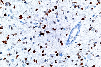
 p53 staining in an anaplastic astrocytoma. | |
| Use | cancer versus benign, prognostication |
| Positive | nuclear staining (>60% or completely negative - see interpretation) |
p53 marks a tumour suppressor protein commonly implicated in cancer and a common immunostain.
Interpretation
- p53 may be one of the most misinterpreted stains.
- TP53 mutations are associated with >60% staining and no staining (0% of cell labelled).[1]
- Reliable interpretation of p53 may require co-assesment of other markers.
See also
References
- ↑ Yemelyanova, A.; Vang, R.; Kshirsagar, M.; Lu, D.; Marks, MA.; Shih, IeM.; Kurman, RJ. (Sep 2011). "Immunohistochemical staining patterns of p53 can serve as a surrogate marker for TP53 mutations in ovarian carcinoma: an immunohistochemical and nucleotide sequencing analysis.". Mod Pathol 24 (9): 1248-53. doi:10.1038/modpathol.2011.85. PMID 21552211.
- ↑ Louis, DN.; Giannini, C.; Capper, D.; Paulus, W.; Figarella-Branger, D.; Lopes, MB.; Batchelor, TT.; Cairncross, JG. et al. (Mar 2018). "cIMPACT-NOW update 2: diagnostic clarifications for diffuse midline glioma, H3 K27M-mutant and diffuse astrocytoma/anaplastic astrocytoma, IDH-mutant.". Acta Neuropathol. doi:10.1007/s00401-018-1826-y. PMID 29497819.