Difference between revisions of "Collecting duct carcinoma"
Jump to navigation
Jump to search
| (5 intermediate revisions by the same user not shown) | |||
| Line 1: | Line 1: | ||
{{ Infobox diagnosis | {{ Infobox diagnosis | ||
| Name = {{PAGENAME}} | | Name = {{PAGENAME}} | ||
| Image = | | Image = Collecting duct carcinoma - 2 WBAL.tif | ||
| Width = | | Width = | ||
| Caption = | | Caption = Collecting duct carcinoma. (WC/George Netto) | ||
| Micro = tubular structures with tapered ends, [[hobnail pattern]], nuclear pleomorphism), high mitotic rate | | Micro = tubular structures with tapered ends, [[hobnail pattern]], nuclear pleomorphism), high mitotic rate | ||
| Subtypes = | | Subtypes = | ||
| LMDDx = [[papillary renal cell carcinoma]] | | LMDDx = [[papillary renal cell carcinoma]], [[urothelial carcinoma]] (with glandular differentiation), metastatic [[adenocarcinoma]], [[renal medullary carcinoma]], [[ALK-rearranged renal cell carcinoma]] | ||
| Stains = | | Stains = | ||
| IHC = CD117 +ve, CK7 +ve, PAX8 +ve, CD10 -ve, AMACR -ve, p63 -ve | | IHC = CD117 +ve, CK7 +ve, PAX8 +ve, CD10 -ve, AMACR -ve, p63 -ve | ||
| Line 54: | Line 54: | ||
DDx: | DDx: | ||
*[[Papillary renal cell carcinoma]] | *[[Papillary renal cell carcinoma]]. | ||
*[[Urothelial carcinoma]] with glandular differentiation. | *[[Urothelial carcinoma]] with glandular differentiation. | ||
*Metastatic [[adenocarcinoma]]. | *Metastatic [[adenocarcinoma]]. | ||
| Line 61: | Line 61: | ||
===Images=== | ===Images=== | ||
<gallery> | |||
Image: Collecting duct carcinoma - 1 WBAL.tif | CDC - 1 (WC/Netto) | |||
Image: Collecting duct carcinoma - 2 WBAL.tif | CDC - 2 (WC/Netto) | |||
*[http:// | Image: Collecting duct carcinoma - 5 WBAL.tif | CDC - 5 (WC/Netto) | ||
</gallery> | |||
====www==== | |||
*[http://images.rsna.org/index.html?doi=10.1148/rg.266065010&fig=F17 CDC (rsna.org)].<ref name=pmid17102051>{{Cite journal | last1 = Prasad | first1 = SR. | last2 = Humphrey | first2 = PA. | last3 = Catena | first3 = JR. | last4 = Narra | first4 = VR. | last5 = Srigley | first5 = JR. | last6 = Cortez | first6 = AD. | last7 = Dalrymple | first7 = NC. | last8 = Chintapalli | first8 = KN. | title = Common and uncommon histologic subtypes of renal cell carcinoma: imaging spectrum with pathologic correlation. | journal = Radiographics | volume = 26 | issue = 6 | pages = 1795-806; discussion 1806-10 | month = | year = | doi = 10.1148/rg.266065010 | PMID = 17102051 }}</ref> | |||
==IHC== | ==IHC== | ||
| Line 74: | Line 77: | ||
*CD10 -ve. | *CD10 -ve. | ||
*AMACR -ve. | *AMACR -ve. | ||
*[[PAX8]] +ve.<ref name=pmid20463571>{{Cite journal | last1 = Albadine | first1 = R. | last2 = Schultz | first2 = L. | last3 = Illei | first3 = P. | last4 = Ertoy | first4 = D. | last5 = Hicks | first5 = J. | last6 = Sharma | first6 = R. | last7 = Epstein | first7 = JI. | last8 = Netto | first8 = GJ. | title = PAX8 (+)/p63 (-) immunostaining pattern in renal collecting duct carcinoma (CDC): a useful immunoprofile in the differential diagnosis of CDC versus urothelial carcinoma of upper urinary tract. | journal = Am J Surg Pathol | volume = 34 | issue = 7 | pages = 965-9 | month = Jul | year = 2010 | doi = 10.1097/PAS.0b013e3181dc5e8a | PMID = 20463571 }}</ref> | |||
UCC: | UCC: | ||
Latest revision as of 15:10, 20 March 2024
| Collecting duct carcinoma | |
|---|---|
| Diagnosis in short | |
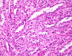
 Collecting duct carcinoma. (WC/George Netto) | |
|
| |
| LM | tubular structures with tapered ends, hobnail pattern, nuclear pleomorphism), high mitotic rate |
| LM DDx | papillary renal cell carcinoma, urothelial carcinoma (with glandular differentiation), metastatic adenocarcinoma, renal medullary carcinoma, ALK-rearranged renal cell carcinoma |
| IHC | CD117 +ve, CK7 +ve, PAX8 +ve, CD10 -ve, AMACR -ve, p63 -ve |
| Gross | medullary tumour |
| Grossing notes | total nephrectomy for tumour grossing, partial nephrectomy grossing |
| Staging | kidney cancer staging |
| Site | kidney - see kidney tumours |
|
| |
| Prevalence | very rare |
| Prognosis | poor |
| Clin. DDx | other renal tumours |
Collecting duct carcinoma, also known as Bellini duct carcinoma[1] and carcinoma of the collecting ducts of Bellini, is a rare aggressive kidney tumour.
It should not be confused with tubulocystic carcinoma of the kidney (also known as low-grade collecting duct carcinoma).
General
- Rare.
- Poor prognosis.
- Usually central location.
- Typically young adults.
Gross
- Medullary location - as opposed to cortical.[2]
Microscopic
Features:[3]
- Tubular structures with tapered ends.
- May be described as tubulopapillary.
- Hobnail pattern - cell width smaller at basement membrane than free surface.[4]
- High grade nuclear features (nuclear pleomorphism).
- High mitotic rate.
Notes:
- Benign urothelium must present to excluded urothelial carcinoma.
- Desmoplastic stroma may be prominent.
DDx:
- Papillary renal cell carcinoma.
- Urothelial carcinoma with glandular differentiation.
- Metastatic adenocarcinoma.
- Renal medullary carcinoma - typically younger, sickle cell trait.
- Hereditary leiomyomatosis and renal cell carcinoma syndrome-associated renal cell carcinoma.
Images
www
IHC
Features:[6]
UCC:
Others:
See also
- Kidney tumours.
- Tubulocystic carcinoma of the kidney (low-grade collecting duct carcinoma).
References
- ↑ Stamatiou, K.; Zizi-Sermpetzoglou, A.. "Bellini duct carcinoma accidentally found upon investigation of uric acid lithiasis.". Indian J Pathol Microbiol 54 (1): 229-30. doi:10.4103/0377-4929.77425. PMID 21393935.
- ↑ López, JI.; Larrinaga, G.; Kuroda, N.; Angulo, JC. (Apr 2015). "The normal and pathologic renal medulla: a comprehensive overview.". Pathol Res Pract 211 (4): 271-80. doi:10.1016/j.prp.2014.12.009. PMID 25595996.
- ↑ Zhou, Ming; Magi-Galluzzi, Cristina (2006). Genitourinary Pathology: A Volume in Foundations in Diagnostic Pathology Series (1st ed.). Churchill Livingstone. pp. 295. ISBN 978-0443066771.
- ↑ Cotran, Ramzi S.; Kumar, Vinay; Fausto, Nelson; Nelso Fausto; Robbins, Stanley L.; Abbas, Abul K. (2005). Robbins and Cotran pathologic basis of disease (7th ed.). St. Louis, Mo: Elsevier Saunders. pp. 1018. ISBN 0-7216-0187-1.
- ↑ Prasad, SR.; Humphrey, PA.; Catena, JR.; Narra, VR.; Srigley, JR.; Cortez, AD.; Dalrymple, NC.; Chintapalli, KN.. "Common and uncommon histologic subtypes of renal cell carcinoma: imaging spectrum with pathologic correlation.". Radiographics 26 (6): 1795-806; discussion 1806-10. doi:10.1148/rg.266065010. PMID 17102051.
- ↑ 6.0 6.1 Srigley, JR.; Delahunt, B. (Jun 2009). "Uncommon and recently described renal carcinomas.". Mod Pathol 22 Suppl 2: S2-S23. doi:10.1038/modpathol.2009.70. PMID 19494850.
- ↑ 7.0 7.1 7.2 Albadine, R.; Schultz, L.; Illei, P.; Ertoy, D.; Hicks, J.; Sharma, R.; Epstein, JI.; Netto, GJ. (Jul 2010). "PAX8 (+)/p63 (-) immunostaining pattern in renal collecting duct carcinoma (CDC): a useful immunoprofile in the differential diagnosis of CDC versus urothelial carcinoma of upper urinary tract.". Am J Surg Pathol 34 (7): 965-9. doi:10.1097/PAS.0b013e3181dc5e8a. PMID 20463571.
- ↑ Kim, MK.; Kim, S. (Dec 2002). "Immunohistochemical profile of common epithelial neoplasms arising in the kidney.". Appl Immunohistochem Mol Morphol 10 (4): 332-8. PMID 12613443.
- ↑ Merino, MJ.; Torres-Cabala, C.; Pinto, P.; Linehan, WM. (Oct 2007). "The morphologic spectrum of kidney tumors in hereditary leiomyomatosis and renal cell carcinoma (HLRCC) syndrome.". Am J Surg Pathol 31 (10): 1578-85. doi:10.1097/PAS.0b013e31804375b8. PMID 17895761.


