Difference between revisions of "Adenosquamous carcinoma of the lung"
Jump to navigation
Jump to search
| (5 intermediate revisions by the same user not shown) | |||
| Line 40: | Line 40: | ||
==Microscopic== | ==Microscopic== | ||
Features: | Features:<ref name=pmid25002356>{{Cite journal | last1 = Rao | first1 = N. | title = Adenosquamous carcinoma. | journal = Semin Diagn Pathol | volume = 31 | issue = 4 | pages = 271-7 | month = Jul | year = 2014 | doi = 10.1053/j.semdp.2014.06.004 | PMID = 25002356 }}</ref><ref name=pmid26068980>{{cite journal |authors=Vassella E, Langsch S, Dettmer MS, Schlup C, Neuenschwander M, Frattini M, Gugger M, Schäfer SC |title=Molecular profiling of lung adenosquamous carcinoma: hybrid or genuine type? |journal=Oncotarget |volume=6 |issue=27 |pages=23905–16 |date=September 2015 |pmid=26068980 |pmc=4695160 |doi=10.18632/oncotarget.4163 |url=}}</ref> | ||
#Adenocarcinoma component. | #Adenocarcinoma component (>=10% of tumour ‡). | ||
#Squamous cell carcinoma component. | #Squamous cell carcinoma component (>=10% of tumour ‡). | ||
Note: | |||
*‡ Criteria used by individual pathologists may vary and/or may not be uniformly applied.<ref name=pmid25002356>{{Cite journal | last1 = Rao | first1 = N. | title = Adenosquamous carcinoma. | journal = Semin Diagn Pathol | volume = 31 | issue = 4 | pages = 271-7 | month = Jul | year = 2014 | doi = 10.1053/j.semdp.2014.06.004 | PMID = 25002356 }}</ref> | |||
DDx: | |||
*[[Squamous cell carcinoma of the lung]]. | |||
*[[Adenocarcinoma of the lung]]. | |||
*Metastatic [[adenosquamous carcinoma]] - requires history. | |||
===Images=== | ===Images=== | ||
<gallery> | <gallery> | ||
Image: Adenosquamous carcinoma of lung -- intermed mag.jpg | ASC | Image: Adenosquamous carcinoma of lung -- intermed mag.jpg | ASC - intermed. mag. (WC) | ||
Image: Adenosquamous carcinoma of lung - alt -- intermed mag.jpg | ASC | Image: Adenosquamous carcinoma of lung - alt -- intermed mag.jpg | ASC - intermed. mag. (WC) | ||
Image: Adenosquamous carcinoma of lung -- high mag.jpg | ASC | Image: Adenosquamous carcinoma of lung -- high mag.jpg | ASC - high mag. (WC) | ||
Image: Adenosquamous carcinoma of lung -- very high mag.jpg | ASC | Image: Adenosquamous carcinoma of lung -- very high mag.jpg | ASC - very high mag. (WC) | ||
</gallery> | </gallery> | ||
====www==== | ====www==== | ||
*[http://www.webpathology.com/image.asp?case=415&n=21 Lung adenosquamous carcinoma (webpathology.com)]. | *[http://www.webpathology.com/image.asp?case=415&n=21 Lung adenosquamous carcinoma (webpathology.com)]. | ||
*[https://www.flickr.com/photos/pulmonary_pathology/7491350926/ Lung adenosquamous carcinoma (flickr.com/Yale Rosen)]. | *[https://www.flickr.com/photos/pulmonary_pathology/7491350926/ Lung adenosquamous carcinoma (flickr.com/Yale Rosen)]. | ||
==Sign out== | |||
===Biopsy - possible adenosquamous carcinoma=== | |||
<pre> | |||
Left Upper Lobe of Lung, Core Biopsy: | |||
- NONSMALL CELL CARCINOMA, FAVOUR ADENOCARCINOMA, CANNOT EXCLUDE ADENOSQUAMOUS CARCINOMA. | |||
Comment: | |||
The tumour stains as follow: | |||
POSITIVE: TTF-1, CK5/6 (focal), p63, napsin A. | |||
NEGATIVE: (none). | |||
The positive staining (TTF-1 and napsin A versus CK5/6 and p63) appears to be in different tumour cells. | |||
The case was reviewed internally and there is agreement on the above interpretation. | |||
</pre> | |||
Note: | |||
*Cases should not be signed as ''adenosquamous carcinoma'' on biopsy, due to the percentage requirements (>=10% for both components) - see ''microscopic'' section. | |||
==See also== | ==See also== | ||
Latest revision as of 14:48, 6 April 2023
| Adenosquamous carcinoma of the lung | |
|---|---|
| Diagnosis in short | |
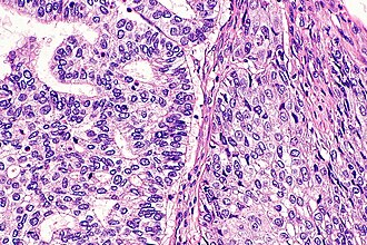
 Adenosquamous carcinoma of lung. H&E stain. | |
| LM DDx | squamous cell carcinoma of the lung, adenocarcinoma of the lung |
| Site | lung - see lung tumours |
|
| |
| Prevalence | rare |
| Clin. DDx | other lung tumours |
Adenosquamous carcinoma of the lung, also lung adenosquamous carcinoma, is a rare epithelial derived malignancy of the lung with features of both adenocarcinoma and squamous cell carcinoma.
It is grouped with the non-small cell lung cancers.
General
- Rare.
- Respond to tyrosine kinase inhibitors.[1][2]
Microscopic
- Adenocarcinoma component (>=10% of tumour ‡).
- Squamous cell carcinoma component (>=10% of tumour ‡).
Note:
- ‡ Criteria used by individual pathologists may vary and/or may not be uniformly applied.[3]
DDx:
- Squamous cell carcinoma of the lung.
- Adenocarcinoma of the lung.
- Metastatic adenosquamous carcinoma - requires history.
Images
www
- Lung adenosquamous carcinoma (webpathology.com).
- Lung adenosquamous carcinoma (flickr.com/Yale Rosen).
Sign out
Biopsy - possible adenosquamous carcinoma
Left Upper Lobe of Lung, Core Biopsy:
     - NONSMALL CELL CARCINOMA, FAVOUR ADENOCARCINOMA, CANNOT EXCLUDE ADENOSQUAMOUS CARCINOMA.

Comment:
The tumour stains as follow:
POSITIVE: TTF-1, CK5/6 (focal), p63, napsin A.
NEGATIVE: (none).

The positive staining (TTF-1 and napsin A versus CK5/6 and p63) appears to be in different tumour cells.

The case was reviewed internally and there is agreement on the above interpretation.
Note:
- Cases should not be signed as adenosquamous carcinoma on biopsy, due to the percentage requirements (>=10% for both components) - see microscopic section.
See also
References
- ↑ Song, Z.; Lin, B.; Shao, L.; Zhang, Y. (Sep 2013). "Therapeutic efficacy of gefitinib and erlotinib in patients with advanced lung adenosquamous carcinoma.". J Chin Med Assoc 76 (9): 481-5. doi:10.1016/j.jcma.2013.05.007. PMID 23769878.
- ↑ Iwanaga, K.; Sueoka-Aragane, N.; Nakamura, T.; Mori, D.; Kimura, S. (2012). "The long-term survival of a patient with adenosquamous lung carcinoma harboring EGFR-activating mutations who was treated with gefitinib.". Intern Med 51 (19): 2771-4. PMID 23037472.
- ↑ 3.0 3.1 Rao, N. (Jul 2014). "Adenosquamous carcinoma.". Semin Diagn Pathol 31 (4): 271-7. doi:10.1053/j.semdp.2014.06.004. PMID 25002356.
- ↑ Vassella E, Langsch S, Dettmer MS, Schlup C, Neuenschwander M, Frattini M, Gugger M, Schäfer SC (September 2015). "Molecular profiling of lung adenosquamous carcinoma: hybrid or genuine type?". Oncotarget 6 (27): 23905–16. doi:10.18632/oncotarget.4163. PMC 4695160. PMID 26068980. https://www.ncbi.nlm.nih.gov/pmc/articles/PMC4695160/.


