Difference between revisions of "Descemet's membrane"
Jump to navigation
Jump to search
| (10 intermediate revisions by the same user not shown) | |||
| Line 1: | Line 1: | ||
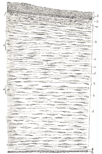
[[Image:Gray871.png|thumb|Drawing of the cornea. Descemet's membrane is a thin layer at the bottom (''4'') of the drawing, between the substantia propria (''3'') and the single layer of endothelial cells (''5''). (WC/Gray's Anatomy)]] | |||
'''Descemet's membrane''' is an [[eye]] pathology [[ditzel]]. | '''Descemet's membrane''' is an [[eye]] pathology [[ditzel]]. | ||
''Descemet's Stripping Automated Endothelial Keratoplasty'', abbreviated ''DSAEK'', redirects to here. | |||
==General== | ==General== | ||
Normal thickness: | |||
*Older individuals | *Younger individuals: 10 +/- 3 micrometres.<ref name=pmid20163865/> | ||
* | *Older individuals: 16 +/- 2 micrometres.<ref name=pmid20163865/> | ||
Note: | |||
*Thickness in ''Fuchs dystrophy'': 34 +/- 11 micrometres.<ref name=pmid20163865>{{Cite journal | last1 = Shousha | first1 = MA. | last2 = Perez | first2 = VL. | last3 = Wang | first3 = J. | last4 = Ide | first4 = T. | last5 = Jiao | first5 = S. | last6 = Chen | first6 = Q. | last7 = Chang | first7 = V. | last8 = Buchser | first8 = N. | last9 = Dubovy | first9 = SR. | title = Use of ultra-high-resolution optical coherence tomography to detect in vivo characteristics of Descemet's membrane in Fuchs' dystrophy. | journal = Ophthalmology | volume = 117 | issue = 6 | pages = 1220-7 | month = Jun | year = 2010 | doi = 10.1016/j.ophtha.2009.10.027 | PMID = 20163865 }}</ref> | |||
==Specific entities== | |||
===Fuchs dystrophy=== | |||
{{Main|Fuchs dystrophy}} | |||
== | ===Pseudophakic bullous keratopathy=== | ||
{{Main|Pseudophakic bullous keratopathy}} | |||
==Sign out== | ==Sign out== | ||
| Line 15: | Line 23: | ||
<pre> | <pre> | ||
Right Eye, Descemet's Membrane, Excision: | Right Eye, Descemet's Membrane, Excision: | ||
- Thin Descemet's membrane | - Thin Descemet's membrane. | ||
- This case will be sent to an eye pathologist for an opinion. | - This case will be sent to an eye pathologist for an opinion. | ||
</pre> | </pre> | ||
==See also== | ==See also== | ||
*[[Eye]]. | *[[Eye]]. | ||
*[[Eyelid]]. | *[[Eyelid]]. | ||
==References== | |||
{{Reflist|1}} | |||
[[Category:Eye]] | [[Category:Eye]] | ||
Latest revision as of 15:44, 13 January 2022
Descemet's membrane is an eye pathology ditzel.
Descemet's Stripping Automated Endothelial Keratoplasty, abbreviated DSAEK, redirects to here.
General
Normal thickness:
Note:
- Thickness in Fuchs dystrophy: 34 +/- 11 micrometres.[1]
Specific entities
Fuchs dystrophy
Main article: Fuchs dystrophy
Pseudophakic bullous keratopathy
Main article: Pseudophakic bullous keratopathy
Sign out
Thin
Right Eye, Descemet's Membrane, Excision:
- Thin Descemet's membrane.
- This case will be sent to an eye pathologist for an opinion.
See also
References
- ↑ 1.0 1.1 1.2 Shousha, MA.; Perez, VL.; Wang, J.; Ide, T.; Jiao, S.; Chen, Q.; Chang, V.; Buchser, N. et al. (Jun 2010). "Use of ultra-high-resolution optical coherence tomography to detect in vivo characteristics of Descemet's membrane in Fuchs' dystrophy.". Ophthalmology 117 (6): 1220-7. doi:10.1016/j.ophtha.2009.10.027. PMID 20163865.
