Difference between revisions of "Ganglion cyst"
(+images) |
|||
| (11 intermediate revisions by the same user not shown) | |||
| Line 7: | Line 7: | ||
| Micro = empty space(s) - usually multiple; fibrotic wall without an epithelial lining +/- myxoid change +/- spindled fibroblasts | | Micro = empty space(s) - usually multiple; fibrotic wall without an epithelial lining +/- myxoid change +/- spindled fibroblasts | ||
| Subtypes = | | Subtypes = | ||
| LMDDx = [[synovial cyst]], [[juxta-articular myxoma]], (other) myxomas, [[digital mucous cyst]] | | LMDDx = [[synovial cyst]], [[juxta-articular myxoma]], (other) myxomas, [[digital mucous cyst]], [[myxoid lesions]] | ||
| Stains = | | Stains = | ||
| IHC = | | IHC = | ||
| Line 28: | Line 28: | ||
| Other = | | Other = | ||
| ClinDDx = | | ClinDDx = | ||
| Tx = | | Tx = surgical excision or conservative management | ||
}} | }} | ||
'''Ganglion cyst''' is a small benign [[ditzel]] classically found on the wrist. | '''Ganglion cyst''' is a small benign [[ditzel]] classically found on the wrist. | ||
| Line 34: | Line 34: | ||
==General== | ==General== | ||
*Very common. | *Very common. | ||
**Most common tumour of the hand.<ref name=emed_gc>URL: [http://emedicine.medscape.com/article/ | **Most common tumour of the hand.<ref name=emed_gc>URL: [http://emedicine.medscape.com/article/1243454-overview http://emedicine.medscape.com/article/1243454-overview]. Accessed on: 8 February 2012.</ref> | ||
*Classically on the wrist.<ref name=pmid17488856>{{Cite journal | last1 = Hasham | first1 = S. | last2 = Burke | first2 = FD. | title = Diagnosis and treatment of swellings in the hand. | journal = Postgrad Med J | volume = 83 | issue = 979 | pages = 296-300 | month = May | year = 2007 | doi = 10.1136/pgmj.2005.043992 | PMID = 17488856 }}</ref> | *Classically on the wrist.<ref name=pmid17488856>{{Cite journal | last1 = Hasham | first1 = S. | last2 = Burke | first2 = FD. | title = Diagnosis and treatment of swellings in the hand. | journal = Postgrad Med J | volume = 83 | issue = 979 | pages = 296-300 | month = May | year = 2007 | doi = 10.1136/pgmj.2005.043992 | PMID = 17488856 }}</ref> | ||
*May be painful.<ref name=Ref_Derm322>{{Ref Derm|322}}</ref> | *May be painful.<ref name=Ref_Derm322>{{Ref Derm|322}}</ref> | ||
*Many (~60%) regress if left alone.<ref name=pmid24967120>{{Cite journal | last1 = Suen | first1 = M. | last2 = Fung | first2 = B. | last3 = Lung | first3 = CP. | title = Treatment of ganglion cysts. | journal = ISRN Orthop | volume = 2013 | issue = | pages = 940615 | month = | year = 2013 | doi = 10.1155/2013/940615 | PMID = 24967120 }}</ref> | |||
==Gross== | ==Gross== | ||
| Line 44: | Line 45: | ||
*Metacarpal boss - degenerative [[arthritis]].<ref name=pmid432327>{{Cite journal | last1 = Cuono | first1 = CB. | last2 = Watson | first2 = HK. | title = The carpal boss: surgical treatment and etiological considerations. | journal = Plast Reconstr Surg | volume = 63 | issue = 1 | pages = 88-93 | month = Jan | year = 1979 | doi = | PMID = 432327 }}</ref> | *Metacarpal boss - degenerative [[arthritis]].<ref name=pmid432327>{{Cite journal | last1 = Cuono | first1 = CB. | last2 = Watson | first2 = HK. | title = The carpal boss: surgical treatment and etiological considerations. | journal = Plast Reconstr Surg | volume = 63 | issue = 1 | pages = 88-93 | month = Jan | year = 1979 | doi = | PMID = 432327 }}</ref> | ||
*[[Giant cell tumour of tendon sheath]]. | *[[Giant cell tumour of tendon sheath]]. | ||
===Image=== | |||
<gallery> | |||
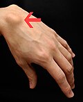
Image: Ganglion-cyst.jpg | Ganglion cyst of the hand. (WC) | |||
</gallery> | |||
==Microscopic== | ==Microscopic== | ||
Features:<ref name=emed_gc>URL: [http://emedicine.medscape.com/article/1243454-overview http://emedicine.medscape.com/article/1243454-overview]. Accessed on: 8 February 2012.</ref><ref name=Ref_Derm322>{{Ref Derm|322}}</ref> | Features:<ref name=emed_gc>URL: [http://emedicine.medscape.com/article/1243454-overview http://emedicine.medscape.com/article/1243454-overview]. Accessed on: 8 February 2012.</ref><ref name=Ref_Derm322>{{Ref Derm|322}}</ref> | ||
| Line 52: | Line 59: | ||
**May have some spindled fibroblasts. | **May have some spindled fibroblasts. | ||
Note: | |||
† The entity is really a pseudocyst.<ref name=emed_gc/> | *† The entity is really a pseudocyst.<ref name=emed_gc/> | ||
DDx: | DDx: | ||
| Line 59: | Line 66: | ||
*[[Juxta-articular myxoma]]. | *[[Juxta-articular myxoma]]. | ||
*Myxoma. | *Myxoma. | ||
*Other [[myxoid lesions]]. | |||
*[[Digital mucous cyst]] - has many spindled fibroblasts, usu. superficial.<ref name=Ref_Derm322>{{Ref Derm|322}}</ref> | *[[Digital mucous cyst]] - has many spindled fibroblasts, usu. superficial.<ref name=Ref_Derm322>{{Ref Derm|322}}</ref> | ||
*[[Nodular hidradenoma]]. | |||
===Images=== | ===Images=== | ||
| Line 71: | Line 80: | ||
www: | www: | ||
*[http://www.surgicalpathologyatlas.com/glfusion/mediagallery/media.php?f=0&sort=0&s=2008080217190580 Ganglion cyst (surgicalpathologyatlas.com)]. | *[http://www.surgicalpathologyatlas.com/glfusion/mediagallery/media.php?f=0&sort=0&s=2008080217190580 Ganglion cyst (surgicalpathologyatlas.com)]. | ||
==Stains== | |||
*Alcian blue +ve (mucin).<ref name=pmid24891856 >{{Cite journal | last1 = Park | first1 = JH. | last2 = Im | first2 = SB. | last3 = Kim | first3 = HK. | last4 = Hwang | first4 = SC. | last5 = Shin | first5 = DS. | last6 = Shin | first6 = WH. | last7 = Kim | first7 = BT. | title = Histopathological findings of hemorrhagic ganglion cyst causing acute radicular pain: a case report. | journal = Korean J Spine | volume = 10 | issue = 4 | pages = 242-5 | month = Dec | year = 2013 | doi = 10.14245/kjs.2013.10.4.242 | PMID = 24891856 | PMC = 4040643}}</ref> | |||
===Images=== | |||
*[http://www.ncbi.nlm.nih.gov/pmc/articles/PMC4040643/figure/F4/ Ganglion cyst - Alican blue staining (nih.gov)].<ref name=pmid24891856/> | |||
==Sign out== | ==Sign out== | ||
<pre> | |||
Soft Tissue (Submitted as "Ganglion from Left Wrist"), Excision: | |||
- Ganglion cyst. | |||
</pre> | |||
===Block letters=== | |||
<pre> | <pre> | ||
SOFT TISSUE ("GANGLION CYST"), LEFT WRIST, EXCISION: | SOFT TISSUE ("GANGLION CYST"), LEFT WRIST, EXCISION: | ||
Latest revision as of 17:49, 12 December 2023
| Ganglion cyst | |
|---|---|
| Diagnosis in short | |
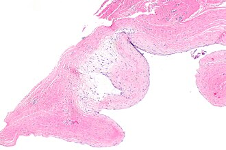
 Ganglion cyst. H&E stain. | |
|
| |
| LM | empty space(s) - usually multiple; fibrotic wall without an epithelial lining +/- myxoid change +/- spindled fibroblasts |
| LM DDx | synovial cyst, juxta-articular myxoma, (other) myxomas, digital mucous cyst, myxoid lesions |
| Site | soft tissue close to joints - esp. in hand |
|
| |
| Prevalence | common |
| Prognosis | benign |
| Treatment | surgical excision or conservative management |
Ganglion cyst is a small benign ditzel classically found on the wrist.
General
- Very common.
- Most common tumour of the hand.[1]
- Classically on the wrist.[2]
- May be painful.[3]
- Many (~60%) regress if left alone.[4]
Gross
- Mass at a joint - classically in the hand.
DDx - clinical:
- Metacarpal boss - degenerative arthritis.[5]
- Giant cell tumour of tendon sheath.
Image
Microscopic
- Empty space(s); usually multiple.
- Fibrotic wall.
- No epithelial lining.†
- +/-Myxoid change - very common.
- May have some spindled fibroblasts.
Note:
- † The entity is really a pseudocyst.[1]
DDx:
- Synovial cyst - true cyst (has an epithelial lining), villous projections into the cystic space.
- Juxta-articular myxoma.
- Myxoma.
- Other myxoid lesions.
- Digital mucous cyst - has many spindled fibroblasts, usu. superficial.[3]
- Nodular hidradenoma.
Images
www:
Stains
- Alcian blue +ve (mucin).[6]
Images
Sign out
Soft Tissue (Submitted as "Ganglion from Left Wrist"), Excision:
- Ganglion cyst.
Block letters
SOFT TISSUE ("GANGLION CYST"), LEFT WRIST, EXCISION:
- GANGLION CYST.
Micro
The sections show fibroadipose tissue with cyst-like spaces surrounded by fibrous tissue. There are no villous projections into the cyst-like spaces. Focal myxoid areas are present with rare bland spindle cells. There is no nuclear atypia and no mitotic activity is appreciated. Dense connective tissue consistent with tendon is present focally.
Alternate
The sections show cyst-like spaces surrounded by fibrous tissue. There are no villous projections into the cyst-like spaces. Focal myxoid areas are present with rare bland spindle cells. There is no nuclear atypia and no mitotic activity is appreciated. Dense connective tissue consistent with tendon is present focally. Benign glands are present.
See also
References
- ↑ 1.0 1.1 1.2 URL: http://emedicine.medscape.com/article/1243454-overview. Accessed on: 8 February 2012.
- ↑ Hasham, S.; Burke, FD. (May 2007). "Diagnosis and treatment of swellings in the hand.". Postgrad Med J 83 (979): 296-300. doi:10.1136/pgmj.2005.043992. PMID 17488856.
- ↑ 3.0 3.1 3.2 Busam, Klaus J. (2009). Dermatopathology: A Volume in the Foundations in Diagnostic Pathology Series (1st ed.). Saunders. pp. 322. ISBN 978-0443066542.
- ↑ Suen, M.; Fung, B.; Lung, CP. (2013). "Treatment of ganglion cysts.". ISRN Orthop 2013: 940615. doi:10.1155/2013/940615. PMID 24967120.
- ↑ Cuono, CB.; Watson, HK. (Jan 1979). "The carpal boss: surgical treatment and etiological considerations.". Plast Reconstr Surg 63 (1): 88-93. PMID 432327.
- ↑ 6.0 6.1 Park, JH.; Im, SB.; Kim, HK.; Hwang, SC.; Shin, DS.; Shin, WH.; Kim, BT. (Dec 2013). "Histopathological findings of hemorrhagic ganglion cyst causing acute radicular pain: a case report.". Korean J Spine 10 (4): 242-5. doi:10.14245/kjs.2013.10.4.242. PMC 4040643. PMID 24891856. https://www.ncbi.nlm.nih.gov/pmc/articles/PMC4040643/.


