Difference between revisions of "Testicular scar"
Jump to navigation
Jump to search
| (4 intermediate revisions by the same user not shown) | |||
| Line 1: | Line 1: | ||
{{ Infobox diagnosis | {{ Infobox diagnosis | ||
| Name = {{PAGENAME}} | | Name = {{PAGENAME}} | ||
| Image = Testicular_scar_--_low_mag.jpg | | Image = Testicular_scar_--_low_mag.jpg | ||
| Width = | | Width = | ||
| Caption = Testicular scar. [[H&E stain]]. | | Caption = Testicular scar. [[H&E stain]]. | ||
| Line 34: | Line 34: | ||
==General== | ==General== | ||
*Well-reported uncommon finding. | *Well-reported uncommon finding. | ||
*May result from the spontaneous regression of a (germ cell) tumour.<ref name=pmid16819328>{{cite journal |author=Balzer BL, Ulbright TM |title=Spontaneous regression of testicular germ cell tumors: an analysis of 42 cases |journal=Am. J. Surg. Pathol. |volume=30 |issue=7 |pages=858–65 |year=2006 |month=July |pmid=16819328 |doi=10.1097/01.pas.0000209831.24230.56 |url=}}</ref> | |||
*Can be the result of trauma, (a prior untreated) [[testicular torsion]] or infection. | |||
==Gross== | ==Gross== | ||
| Line 49: | Line 51: | ||
*[[necrosis|Necrotic]] tumour. | *[[necrosis|Necrotic]] tumour. | ||
*[[Germ cell tumour]], e.g. [[seminoma]]. | *[[Germ cell tumour]], e.g. [[seminoma]]. | ||
*Regressed germ cell tumour - suggestive features:<ref name=pmid16819328/> | |||
**Distinct scar +/-[[germ cell neoplasia in situ]] (GCNIS) +/-coarse calcifications (intratubular). | |||
**Scar with prominent (small) blood vessels (weakly suggestive). | |||
**Scar with lymphoplasmacytic inflammatory infiltrate (weakly suggestive). | |||
===Images=== | ===Images=== | ||
Latest revision as of 23:37, 15 March 2016
| Testicular scar | |
|---|---|
| Diagnosis in short | |
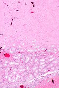
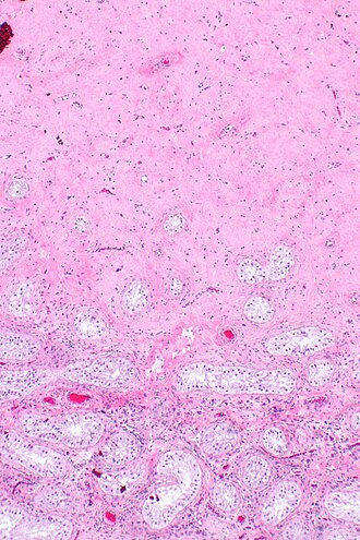 Testicular scar. H&E stain. | |
|
| |
| Synonyms | scar of testis |
|
| |
| LM | seminiferous tubules replaced by fibrosis (hyaline material with a relatively low cellularity and no nuclear atypia) +/-hemosiderin-laden macrophages, atrophic changes (see testicular atrophy) |
| LM DDx | necrotic tumour, (residual) testicular tumour (e.g. germ cell tumour) |
| Gross | tan brown or white, well-circumscribed |
| Site | testis |
|
| |
| Clinical history | +/-history of testicular tumour |
| Prevalence | uncommon |
| Blood work | unremarkable |
| Prognosis | benign |
| Clin. DDx | (residual) testicular tumour |
Testicular scar, also scar of testis, is a phenomenon that may arise in the context of treatment for a germ cell tumour[1] or result from the spontaneous regression of a (germ cell) tumour.[2]
General
- Well-reported uncommon finding.
- May result from the spontaneous regression of a (germ cell) tumour.[2]
- Can be the result of trauma, (a prior untreated) testicular torsion or infection.
Gross
- Tan-brown or white lesion.
- Well-circumscribed.
Microscopic
Features:
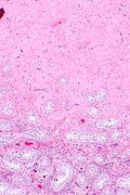
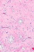
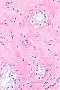
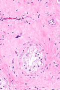
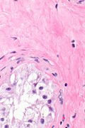
- Seminiferous tubules replaced by fibrosis.
- Hyaline material with a relatively low cellularity and no nuclear atypia.
- +/-Hemosiderin-laden macrophages.
- Atrophic changes[2] - see testicular atrophy.
DDx:
- Necrotic tumour.
- Germ cell tumour, e.g. seminoma.
- Regressed germ cell tumour - suggestive features:[2]
- Distinct scar +/-germ cell neoplasia in situ (GCNIS) +/-coarse calcifications (intratubular).
- Scar with prominent (small) blood vessels (weakly suggestive).
- Scar with lymphoplasmacytic inflammatory infiltrate (weakly suggestive).
Images
Sign out
TESTICLE, RIGHT, ORCHIECTOMY:
- TESTICULAR SCAR REPLACING MANY OF THE SEMINIFEROUS TUBULES.
- REMAINING SEMINIFEROUS TUBULES WITH ATROPHIC CHANGES.
- BENIGN SPERMATIC CORD AND EPIDIDYMIS.
- NO EVIDENCE OF RESIDUAL GERM CELL TUMOUR.
- NEGATIVE FOR MALIGNANCY.
See also
References
- ↑ Ramsey S, Kerr G, Howard GC, Donat R (2013). "Orchidectomy after primary chemotherapy for metastatic testicular cancer". Urol. Int. 91 (4): 439–44. doi:10.1159/000350858. PMID 24021555.
- ↑ 2.0 2.1 2.2 2.3 Balzer BL, Ulbright TM (July 2006). "Spontaneous regression of testicular germ cell tumors: an analysis of 42 cases". Am. J. Surg. Pathol. 30 (7): 858–65. doi:10.1097/01.pas.0000209831.24230.56. PMID 16819328.
