Difference between revisions of "Intestinal metaplasia of the gallbladder"
Jump to navigation
Jump to search
| (14 intermediate revisions by the same user not shown) | |||
| Line 4: | Line 4: | ||
| Width = | | Width = | ||
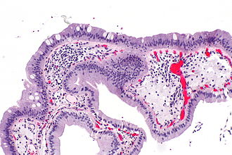
| Caption = Gallbladder with intestinal metaplasia. [[H&E stain]]. | | Caption = Gallbladder with intestinal metaplasia. [[H&E stain]]. | ||
| Micro = | | Micro = gallbladder mucosa with goblet cells +/-Paneth cells | ||
| Subtypes = | | Subtypes = | ||
| LMDDx = gallbladder | | LMDDx = [[gallbladder dysplasia]], [[gallbladder carcinoma]] | ||
| Stains = [[Alcian blue stain]] +ve | | Stains = [[Alcian blue stain]] +ve | ||
| IHC = | | IHC = | ||
| Line 12: | Line 12: | ||
| Molecular = | | Molecular = | ||
| IF = | | IF = | ||
| Gross = | | Gross = | ||
| Grossing = | | Grossing = [[Gallbladder grossing]] | ||
| Site = [[gallbladder]] | | Site = [[gallbladder]] | ||
| Assdx = [[chronic cholecystitis]] | | Assdx = [[chronic cholecystitis]] | ||
| Line 24: | Line 24: | ||
| Rads = | | Rads = | ||
| Endoscopy = | | Endoscopy = | ||
| Prognosis = benign, increased risk of carcinoma | | Prognosis = benign, increased risk of [[gallbladder carcinoma|carcinoma]] | ||
| Other = | | Other = | ||
| ClinDDx = | | ClinDDx = | ||
| Line 41: | Line 41: | ||
==Gross== | ==Gross== | ||
*No specific changes. | *No specific changes. | ||
Notes: | |||
*Usually seen in the context of chronic cholecystitis if isolated. | |||
==Microscopic== | ==Microscopic== | ||
Features:<ref name=pmid2872152>{{Cite journal | last1 = Albores-Saavedra | first1 = J. | last2 = Nadji | first2 = M. | last3 = Henson | first3 = DE. | last4 = Ziegels-Weissman | first4 = J. | last5 = Mones | first5 = JM. | title = Intestinal metaplasia of the gallbladder: a morphologic and immunocytochemical study. | journal = Hum Pathol | volume = 17 | issue = 6 | pages = 614-20 | month = Jun | year = 1986 | doi = | PMID = 2872152 }}</ref> | Features:<ref name=pmid2872152>{{Cite journal | last1 = Albores-Saavedra | first1 = J. | last2 = Nadji | first2 = M. | last3 = Henson | first3 = DE. | last4 = Ziegels-Weissman | first4 = J. | last5 = Mones | first5 = JM. | title = Intestinal metaplasia of the gallbladder: a morphologic and immunocytochemical study. | journal = Hum Pathol | volume = 17 | issue = 6 | pages = 614-20 | month = Jun | year = 1986 | doi = | PMID = 2872152 }}</ref> | ||
*[[Goblet cell]]s - '''key feature'''. | *[[Goblet cell]]s - '''key feature'''. | ||
*+/-Paneth | *+/-[[Paneth cell]]s.<ref name=Ref_Sternberg4_1789>{{Ref Sternberg4|1789}}</ref> | ||
Notes: | |||
*Often accompanied by antral type metplasia. | *Often accompanied by antral type metplasia. | ||
**Gastric antral-type epithelium - may form glands. | **Gastric antral-type epithelium - may form glands. | ||
*Gallbladders with intestinal metaplasia do ''not'' have to be [[submitted in total]].<ref name=uscap2017_akki>Akki ''et al.'' (2017) "Detecting Incidental Gallbladder Adenocarcinoma: When to Submit the Entire Gallbladder". Available at: [http://www.abstracts2view.com/uscap17/view.php?nu=USCAP17L_2016 http://www.abstracts2view.com/uscap17/view.php?nu=USCAP17L_2016]. United States and Canadian Academy of Pathology Annual Meeting. Accessed on: April 9, 2017.</ref> | |||
**Adsay ''et al.'' suggests submitting two additional cassettes of tissue.<ref name=pmid23897266>{{cite journal |authors=Adsay V, Saka B, Basturk O, Roa JC |title=Criteria for pathologic sampling of gallbladder specimens |journal=Am J Clin Pathol |volume=140 |issue=2 |pages=278–80 |date=August 2013 |pmid=23897266 |doi=10.1309/AJCPUJPGQIZ6DC6A |url=}}</ref> | |||
DDx: | DDx: | ||
*[[Gallbladder adenocarcinoma]]. | *[[Gallbladder adenocarcinoma]]. | ||
*[[Gallbladder adenoma]] (gallbladder dysplasia). | |||
===Images=== | ===Images=== | ||
| Line 67: | Line 73: | ||
==Sign out== | ==Sign out== | ||
<pre> | |||
Gallbladder, Cholecystectomy: | |||
- Gallbladder wall with focal intestinal metaplasia and mild chronic inflammation. | |||
- Cholelithiasis. | |||
- NEGATIVE for dysplasia. | |||
</pre> | |||
===Block letters=== | |||
<pre> | <pre> | ||
GALLBLADDER, CHOLECYSTECTOMY: | GALLBLADDER, CHOLECYSTECTOMY: | ||
| Line 74: | Line 88: | ||
- NEGATIVE FOR DYSPLASIA. | - NEGATIVE FOR DYSPLASIA. | ||
</pre> | </pre> | ||
===Micro=== | |||
The sections show gallbladder wall with mild chronic inflammation and entrapped epithelial | |||
crypts, that focally extend through the muscularis. The entrapped deep crypts have no | |||
apparent nuclear atypia, and no apparent mitotic activity. | |||
A very small focus of intestinal metaplasia is present in the surface epithelium. No dysplasia is identified. | |||
==See also== | ==See also== | ||
Latest revision as of 16:22, 9 February 2021
| Intestinal metaplasia of the gallbladder | |
|---|---|
| Diagnosis in short | |
 Gallbladder with intestinal metaplasia. H&E stain. | |
|
| |
| LM | gallbladder mucosa with goblet cells +/-Paneth cells |
| LM DDx | gallbladder dysplasia, gallbladder carcinoma |
| Stains | Alcian blue stain +ve |
| Grossing notes | Gallbladder grossing |
| Site | gallbladder |
|
| |
| Associated Dx | chronic cholecystitis |
| Prognosis | benign, increased risk of carcinoma |
Intestinal metaplasia of the gallbladder is a pathology of the gallbladder associated with an increased risk of gallbladder carcinoma.
It is also known as gallbladder intestinal metaplasia.
General
- Associated with chronic cholecystitis.[1]
- Not associated with acute cholecystitis.
Significance:
- Increased risk of carcinoma.[2]
Gross
- No specific changes.
Notes:
- Usually seen in the context of chronic cholecystitis if isolated.
Microscopic
Features:[3]
- Goblet cells - key feature.
- +/-Paneth cells.[4]
Notes:
- Often accompanied by antral type metplasia.
- Gastric antral-type epithelium - may form glands.
- Gallbladders with intestinal metaplasia do not have to be submitted in total.[5]
- Adsay et al. suggests submitting two additional cassettes of tissue.[6]
DDx:
- Gallbladder adenocarcinoma.
- Gallbladder adenoma (gallbladder dysplasia).
Images
www:
Stains
- Alcian blue/PAS +ve.
Sign out
Gallbladder, Cholecystectomy:
	- Gallbladder wall with focal intestinal metaplasia and mild chronic inflammation.
	- Cholelithiasis.
	- NEGATIVE for dysplasia.
Block letters
GALLBLADDER, CHOLECYSTECTOMY:
- INTESTINAL METAPLASIA OF THE GALLBLADDER, FOCAL.
- CHRONIC CHOLECYSTITIS.
- CHOLELITHIASIS.
- NEGATIVE FOR DYSPLASIA.
Micro
The sections show gallbladder wall with mild chronic inflammation and entrapped epithelial crypts, that focally extend through the muscularis. The entrapped deep crypts have no apparent nuclear atypia, and no apparent mitotic activity.
A very small focus of intestinal metaplasia is present in the surface epithelium. No dysplasia is identified.
See also
References
- ↑ Sai, K.; Kajiwara, H. (2001). "An immunohistochemical study of metaplastic endocrine cells in human gallbladder cancer.". J Hepatobiliary Pancreat Surg 8 (5): 453-60. PMID 11702256.
- ↑ Duarte, I.; Llanos, O.; Domke, H.; Harz, C.; Valdivieso, V. (Sep 1993). "Metaplasia and precursor lesions of gallbladder carcinoma. Frequency, distribution, and probability of detection in routine histologic samples.". Cancer 72 (6): 1878-84. PMID 8364865.
- ↑ Albores-Saavedra, J.; Nadji, M.; Henson, DE.; Ziegels-Weissman, J.; Mones, JM. (Jun 1986). "Intestinal metaplasia of the gallbladder: a morphologic and immunocytochemical study.". Hum Pathol 17 (6): 614-20. PMID 2872152.
- ↑ Mills, Stacey E; Carter, Darryl; Greenson, Joel K; Oberman, Harold A; Reuter, Victor E (2004). Sternberg's Diagnostic Surgical Pathology (4th ed.). Lippincott Williams & Wilkins. pp. 1789. ISBN 978-0781740517.
- ↑ Akki et al. (2017) "Detecting Incidental Gallbladder Adenocarcinoma: When to Submit the Entire Gallbladder". Available at: http://www.abstracts2view.com/uscap17/view.php?nu=USCAP17L_2016. United States and Canadian Academy of Pathology Annual Meeting. Accessed on: April 9, 2017.
- ↑ Adsay V, Saka B, Basturk O, Roa JC (August 2013). "Criteria for pathologic sampling of gallbladder specimens". Am J Clin Pathol 140 (2): 278–80. doi:10.1309/AJCPUJPGQIZ6DC6A. PMID 23897266.
- ↑ Mukhopadhyay, S.; Landas, SK. (Mar 2005). "Putative precursors of gallbladder dysplasia: a review of 400 routinely resected specimens.". Arch Pathol Lab Med 129 (3): 386-90. doi:10.1043/1543-2165(2005)129386:PPOGDA2.0.CO;2. PMID 15737036.


