Difference between revisions of "Rhabdomyoma"
Jump to navigation
Jump to search
| (4 intermediate revisions by the same user not shown) | |||
| Line 1: | Line 1: | ||
{{ Infobox diagnosis | |||
| Name = {{PAGENAME}} | |||
| Image = Rhabdomyoma -- high mag.jpg | |||
| Width = | |||
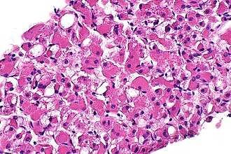
| Caption = Rhabdomyoma. [[H&E stain]]. (WC) | |||
| Synonyms = | |||
| Micro = spider cells (large polygonal cells (~10-20x RBC diameter), abundant cytoplasm with clearing) | |||
| Subtypes = fetal rhabdomyoma, adult rhabdomyoma | |||
| LMDDx = [[granular cell tumour]], [[hibernoma]], [[oncocytoma]] | |||
| Stains = | |||
| IHC = desmin +ve, actin +ve, myoglobin +ve, S-100 -ve | |||
| EM = | |||
| Molecular = | |||
| IF = | |||
| Gross = | |||
| Grossing = | |||
| Staging = | |||
| Site = [[heart]] (see ''[[cardiac tumours]]), [[head and neck pathology|head and neck]] | |||
| Assdx = | |||
| Syndromes = | |||
| Clinicalhx = | |||
| Signs = | |||
| Symptoms = | |||
| Prevalence = rare | |||
| Bloodwork = | |||
| Rads = | |||
| Endoscopy = | |||
| Prognosis = benign | |||
| Other = | |||
| ClinDDx = | |||
| Tx = | |||
}} | |||
'''Rhabdomyoma''' a benign muscle tumour. Often seen in the context of [[tuberous sclerosis]]. | '''Rhabdomyoma''' a benign muscle tumour. Often seen in the context of [[tuberous sclerosis]]. | ||
| Line 22: | Line 54: | ||
DDx: | DDx: | ||
*[[Hibernoma]]. | |||
*[[Granular cell tumour]]. | |||
*[[Oncocytoma]]. | |||
*[[Lipoma]]. | *[[Lipoma]]. | ||
*[[Fat necrosis]]. | *[[Fat necrosis]]. | ||
===Images=== | ===Images=== | ||
| Line 44: | Line 77: | ||
==IHC== | ==IHC== | ||
Features:<ref name=pmid16133368 >{{Cite journal | last1 = Hansen | first1 = T. | last2 = Katenkamp | first2 = D. | title = Rhabdomyoma of the head and neck: morphology and differential diagnosis. | journal = Virchows Arch | volume = 447 | issue = 5 | pages = 849-54 | month = Nov | year = 2005 | doi = 10.1007/s00428-005-0038-8 | PMID = 16133368 }}</ref> | Features:<ref name=pmid16133368 >{{Cite journal | last1 = Hansen | first1 = T. | last2 = Katenkamp | first2 = D. | title = Rhabdomyoma of the head and neck: morphology and differential diagnosis. | journal = Virchows Arch | volume = 447 | issue = 5 | pages = 849-54 | month = Nov | year = 2005 | doi = 10.1007/s00428-005-0038-8 | PMID = 16133368 }}</ref> | ||
*Desmin +ve. | *Desmin +ve (21 of 21 adult cases<ref name=pmid8505039>{{Cite journal | last1 = Kapadia | first1 = SB. | last2 = Meis | first2 = JM. | last3 = Frisman | first3 = DM. | last4 = Ellis | first4 = GL. | last5 = Heffner | first5 = DK. | last6 = Hyams | first6 = VJ. | title = Adult rhabdomyoma of the head and neck: a clinicopathologic and immunophenotypic study. | journal = Hum Pathol | volume = 24 | issue = 6 | pages = 608-17 | month = Jun | year = 1993 | doi = | PMID = 8505039 }}</ref>). | ||
*Myogoblin +ve. | *Myogoblin +ve. | ||
*Actin +ve. | *Actin +ve (21 of 21 adult cases<ref name=pmid8505039/>). | ||
*Vimentin -ve/+ve. | |||
*S-100 -ve. | *S-100 -ve. | ||
**Positive in granular cell tumour and | **Positive in granular cell tumour and hibernoma. | ||
==See also== | ==See also== | ||
*[[Cardiac tumours]]. | *[[Cardiac tumours]]. | ||
*[[Head and neck pathology]]. | |||
==References== | ==References== | ||
Latest revision as of 15:47, 22 April 2024
| Rhabdomyoma | |
|---|---|
| Diagnosis in short | |
 Rhabdomyoma. H&E stain. (WC) | |
|
| |
| LM | spider cells (large polygonal cells (~10-20x RBC diameter), abundant cytoplasm with clearing) |
| Subtypes | fetal rhabdomyoma, adult rhabdomyoma |
| LM DDx | granular cell tumour, hibernoma, oncocytoma |
| IHC | desmin +ve, actin +ve, myoglobin +ve, S-100 -ve |
| Site | heart (see cardiac tumours), head and neck |
|
| |
| Prevalence | rare |
| Prognosis | benign |
Rhabdomyoma a benign muscle tumour. Often seen in the context of tuberous sclerosis.
General
- May be seen in the context of tuberous sclerosis.
- Rare benign mesenchymal tumour - may be seen in the head and neck.[1]
- Can cause death if in the heart.[2]
Gross
- Solid, white/tan colour.
Image:
Microscopic
Features - cardiac:[3]
- Spider cells:
- Large polygonal cells (~10-20x RBC diameter):
- Abundant cytoplasm filled with glycogen.
- Large polygonal cells (~10-20x RBC diameter):
Note:
- Fetal rhabdomyomas may have pseudoepitheliomatous hyperplasia.[1]
DDx:
Images
www
IHC
Features:[1]
- Desmin +ve (21 of 21 adult cases[4]).
- Myogoblin +ve.
- Actin +ve (21 of 21 adult cases[4]).
- Vimentin -ve/+ve.
- S-100 -ve.
- Positive in granular cell tumour and hibernoma.
See also
References
- ↑ 1.0 1.1 1.2 Hansen, T.; Katenkamp, D. (Nov 2005). "Rhabdomyoma of the head and neck: morphology and differential diagnosis.". Virchows Arch 447 (5): 849-54. doi:10.1007/s00428-005-0038-8. PMID 16133368.
- ↑ Neri, M.; Di Donato, S.; Maglietta, R.; Pomara, C.; Riezzo, I.; Turillazzi, E.; Fineschi, V. (Dec 2012). "Sudden death as presenting symptom caused by cardiac primary multicentric left ventricle rhabdomyoma, in an 11-month-old baby. An immunohistochemical study.". Diagn Pathol 7: 169. doi:10.1186/1746-1596-7-169. PMID 23206573.
- ↑ URL: http://www.brown.edu/Courses/Digital_Path/systemic_path/cardio/rhabdomyoma.html. Accessed on: 19 October 2011.
- ↑ 4.0 4.1 Kapadia, SB.; Meis, JM.; Frisman, DM.; Ellis, GL.; Heffner, DK.; Hyams, VJ. (Jun 1993). "Adult rhabdomyoma of the head and neck: a clinicopathologic and immunophenotypic study.". Hum Pathol 24 (6): 608-17. PMID 8505039.


