Difference between revisions of "Chalazion"
Jump to navigation
Jump to search
(split out) |
|||
| (One intermediate revision by the same user not shown) | |||
| Line 39: | Line 39: | ||
==Sign out== | ==Sign out== | ||
<pre> | |||
Eyelid Lesion, Right Upper, Biopsy: | |||
- Lipogranulomas with active inflammation and lymphoplasmacytic infiltrate. | |||
- Reactive mucosa. | |||
Comment: | |||
The findings are consistent with a chalazion. | |||
</pre> | |||
===Block letters=== | |||
<pre> | <pre> | ||
EYELID LESION, RIGHT UPPER, BIOPSY: | EYELID LESION, RIGHT UPPER, BIOPSY: | ||
| Line 68: | Line 78: | ||
==See also== | ==See also== | ||
*[[Eye]]. | *[[Eye]]. | ||
*[[Eyelid]]. | |||
==References== | ==References== | ||
Latest revision as of 17:35, 13 December 2021
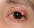
The chalazion is a common benign pathology of the eyelid.
General
- Benign eye thingy that arises from the special sebaceous gland associated with the eyelid (Meibomian gland).
- Usually diagnosed based on clinical appearance - accuracy ~94% in one series.[1]
Gross
- Focal eyelid swelling - typically upper eyelid.
DDx (clinical):[1]
Images
www:
Microscopic
Features:
- Lipogranulomas - key feature.[1]
- Granulomatous inflammation around clear spaces (lipid).[2]
- Multinucleated giant cells - common.
- Plasma cells - abundant - important.
DDx:[4]
Images
www:
Sign out
Eyelid Lesion, Right Upper, Biopsy:
	- Lipogranulomas with active inflammation and lymphoplasmacytic infiltrate.
	- Reactive mucosa.

Comment:
The findings are consistent with a chalazion.
Block letters
EYELID LESION, RIGHT UPPER, BIOPSY:
- LIPOGRANULOMAS.
- LYMPHOPLASMACYTIC RICH INFLAMMATORY INFILTRATE.
- GRANULATION TISSUE.
- REACTIVE SQUAMOUS MUCOSA.

COMMENT:
The findings are consistent with a chalazion.

Special stains (ZN, PASD, GMS) did not demonstrate microorganisms.
Alternate
SKIN LESION, LEFT LOWER EYELID, PUNCH BIOPSY:
- LIPOGRANULOMAS WITH GIANT CELLS AND A LYMPHOPLASMACYTIC RICH INFLAMMATORY
  INFILTRATE -- CONSISTENT WITH CHALAZION.
- GRANULATION TISSUE.
- NEGATIVE FOR MALIGNANCY.
Micro
The sections show a reactive squamous mucosa with palisading granulomas that surround clear spaces (lipid). This is accompanied by a lymphoplasmacytic rich inflammatory infiltrate. Granulation tissue is present. Rare multinucleated giant cells are identified. Neutrophils are numerous and seen in association with the histiocytes.
No significant nuclear atypia is apparent.
See also
References
- ↑ 1.0 1.1 1.2 1.3 Ozdal, PC.; Codère, F.; Callejo, S.; Caissie, AL.; Burnier, MN. (Feb 2004). "Accuracy of the clinical diagnosis of chalazion.". Eye (Lond) 18 (2): 135-8. doi:10.1038/sj.eye.6700603. PMID 14762403. http://www.nature.com/eye/journal/v18/n2/full/6700603a.html.
- ↑ D'hermies, F.; Fayet, B.; Meyer, A.; Morel, X.; Halhal, M.; Elmaleh, C.; Azan, F.; Behar-Cohen, F. et al. (Feb 2004). "[Chalazion mimicking an eyelid tumor].". J Fr Ophtalmol 27 (2): 202-5. PMID 15029054.
- ↑ URL: http://emedicine.medscape.com/article/1212709-workup. Accessed on: 9 February 2012.
- ↑ Tadrous, Paul.J. Diagnostic Criteria Handbook in Histopathology: A Surgical Pathology Vade Mecum (1st ed.). Wiley. pp. 194. ISBN 978-0470519035.

