Difference between revisions of "Angiodysplasia"
Jump to navigation
Jump to search
(→Gross) |
|||
| (7 intermediate revisions by the same user not shown) | |||
| Line 1: | Line 1: | ||
{{ Infobox diagnosis | |||
| Name = {{PAGENAME}} | |||
| Image = Compatible with angiodysplasia -- low mag.jpg | |||
| Width = | |||
| Caption = Compatible with angiodysplasia. [[H&E stain]]. | |||
| Synonyms = | |||
| Micro = dilated vessels in mucosa and submucosa | |||
| Subtypes = | |||
| LMDDx = prominent vessels | |||
| Stains = | |||
| IHC = | |||
| EM = | |||
| Molecular = | |||
| IF = | |||
| Gross = | |||
| Grossing = | |||
| Staging = | |||
| Site = | |||
| Assdx = | |||
| Syndromes = | |||
| Clinicalhx = older individuals | |||
| Signs = bleeding from rectum | |||
| Symptoms = | |||
| Prevalence = | |||
| Bloodwork = | |||
| Rads = | |||
| Endoscopy = red lesion - typically right colon or cecum | |||
| Prognosis = | |||
| Other = | |||
| ClinDDx = Other causes of [[Colon#Bleeding|lower GI bleed]] | |||
| Tx = | |||
}} | |||
'''Angiodysplasia''' is a benign pathology of the large bowel. | '''Angiodysplasia''' is a benign pathology of the large bowel. | ||
==General== | ==General== | ||
*[[Colon#Bleeding| | *[[Clinical diagnosis]]. | ||
*[[Colon#Bleeding|Cause of (lower) GI haemorrhage]]. | |||
*Generally, not a problem pathologists see. | *Generally, not a problem pathologists see. | ||
*May be associated with aortic stenosis; known as ''Heyde syndrome''.<ref name=pmid19652242>{{cite journal |author=Hui YT, Lam WM, Fong NM, Yuen PK, Lam JT |title=Heyde's syndrome: diagnosis and management by the novel single-balloon enteroscopy |journal=Hong Kong Med J |volume=15 |issue=4 |pages=301–3 |year=2009 |month=August |pmid=19652242 |doi= |url=http://www.hkmj.org/abstracts/v15n4/301.htm}}</ref> | *May be associated with aortic stenosis; known as ''Heyde syndrome''.<ref name=pmid19652242>{{cite journal |author=Hui YT, Lam WM, Fong NM, Yuen PK, Lam JT |title=Heyde's syndrome: diagnosis and management by the novel single-balloon enteroscopy |journal=Hong Kong Med J |volume=15 |issue=4 |pages=301–3 |year=2009 |month=August |pmid=19652242 |doi= |url=http://www.hkmj.org/abstracts/v15n4/301.htm}}</ref> | ||
| Line 17: | Line 50: | ||
Note: | Note: | ||
*[[Crohn's disease]] - may mimic angiodysplasia radiographically.<ref name=pmid3054852/> | *[[Crohn's disease]] - may mimic angiodysplasia radiographically.<ref name=pmid3054852/> | ||
===Images=== | |||
<gallery> | |||
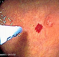
Image:Argon plasma coagulation.jpg | Angiodysplasia - endoscopy. (WC/Grover) | |||
</gallery> | |||
==Microscopic== | ==Microscopic== | ||
Features:<ref name=pmid3054852>{{Cite journal | last1 = Hemingway | first1 = AP. | title = Angiodysplasia: current concepts. | journal = Postgrad Med J | volume = 64 | issue = 750 | pages = 259-63 | month = Apr | year = 1988 | doi = | PMID = 3054852 }}</ref> | Features:<ref name=pmid3054852>{{Cite journal | last1 = Hemingway | first1 = AP. | title = Angiodysplasia: current concepts. | journal = Postgrad Med J | volume = 64 | issue = 750 | pages = 259-63 | month = Apr | year = 1988 | doi = | PMID = 3054852 }}</ref> | ||
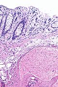
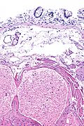
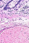
*Dilated vessels in mucosa and submucosa. | *Dilated [[blood vessels]] in the mucosa and submucosa. | ||
DDx: | |||
*[[Crohn's disease]] - may be associated increase vascularity. | |||
===Images=== | |||
<gallery> | |||
Image: Compatible with angiodysplasia -- extremely low mag.jpg | AD - extremely low mag. | |||
Image: Compatible with angiodysplasia -- very low mag.jpg | AD - very low mag. | |||
Image: Compatible with angiodysplasia -- low mag.jpg | AD - low mag. | |||
Image: Compatible with angiodysplasia -- intermed mag.jpg | AD - intermed. mag. | |||
Image: Compatible with angiodysplasia - alt -- intermed mag.jpg | AD - intermed. mag. | |||
Image: Compatible with angiodysplasia -- high mag.jpg | AD - high mag. | |||
</gallery> | |||
==See also== | ==See also== | ||
Latest revision as of 03:10, 23 November 2016
| Angiodysplasia | |
|---|---|
| Diagnosis in short | |
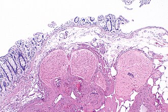 Compatible with angiodysplasia. H&E stain. | |
|
| |
| LM | dilated vessels in mucosa and submucosa |
| LM DDx | prominent vessels |
| Clinical history | older individuals |
| Signs | bleeding from rectum |
| Endoscopy | red lesion - typically right colon or cecum |
| Clin. DDx | Other causes of lower GI bleed |
Angiodysplasia is a benign pathology of the large bowel.
General
- Clinical diagnosis.
- Cause of (lower) GI haemorrhage.
- Generally, not a problem pathologists see.
- May be associated with aortic stenosis; known as Heyde syndrome.[1]
Epidemiology:
- Older people.
Etiology:
- Thought to be caused by the higher wall tension of cecum (due to larger diameter) and result from (intermittent) venous occlusion/focal dilation of vessels.[2]
Gross
- Cecum - classic location.
Note:
- Crohn's disease - may mimic angiodysplasia radiographically.[3]
Images
Microscopic
Features:[3]
- Dilated blood vessels in the mucosa and submucosa.
DDx:
- Crohn's disease - may be associated increase vascularity.
Images
See also
References
- ↑ Hui YT, Lam WM, Fong NM, Yuen PK, Lam JT (August 2009). "Heyde's syndrome: diagnosis and management by the novel single-balloon enteroscopy". Hong Kong Med J 15 (4): 301–3. PMID 19652242. http://www.hkmj.org/abstracts/v15n4/301.htm.
- ↑ Cotran, Ramzi S.; Kumar, Vinay; Fausto, Nelson; Nelso Fausto; Robbins, Stanley L.; Abbas, Abul K. (2005). Robbins and Cotran pathologic basis of disease (7th ed.). St. Louis, Mo: Elsevier Saunders. pp. 854. ISBN 0-7216-0187-1.
- ↑ 3.0 3.1 Hemingway, AP. (Apr 1988). "Angiodysplasia: current concepts.". Postgrad Med J 64 (750): 259-63. PMID 3054852.
