Difference between revisions of "Cholelithiasis"
Jump to navigation
Jump to search
| (5 intermediate revisions by the same user not shown) | |||
| Line 1: | Line 1: | ||
{{ Infobox diagnosis | |||
| Name = {{PAGENAME}} | |||
| Image = Gallensteine_2006_03_28.JPG | |||
| Width = | |||
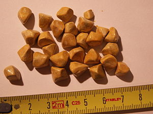
| Caption = Yellow gallstones. (WC) | |||
| Synonyms = | |||
| Micro = | |||
| Subtypes = cholesterol stones, pigment stones, mixed cholesterol/pigment stones | |||
| LMDDx = | |||
| Stains = | |||
| IHC = | |||
| EM = | |||
| Molecular = | |||
| IF = | |||
| Gross = yellow, black/brown or mixed (dependent on subtype) | |||
| Grossing = [[gallbladder grossing]] | |||
| Staging = | |||
| Site = [[gallbladder]] | |||
| Assdx = choledocholithiasis, (gallstone) [[pancreatitis]], [[acute cholecystitis]] (esp. if in gallbladder neck), [[chronic cholecystitis]] | |||
| Syndromes = | |||
| Clinicalhx = | |||
| Signs = | |||
| Symptoms = | |||
| Prevalence = very common | |||
| Bloodwork = | |||
| Rads = | |||
| Endoscopy = | |||
| Prognosis = benign | |||
| Other = | |||
| ClinDDx = | |||
| Tx = | |||
}} | |||
'''Cholelithiasis''' is the formation of stones (gallstones) in the [[gallbladder]]. | '''Cholelithiasis''' is the formation of stones (gallstones) in the [[gallbladder]]. | ||
| Line 45: | Line 77: | ||
==Microscopic== | ==Microscopic== | ||
*Not routinely done on gallstones. | *'''Not''' routinely done on gallstones. | ||
===Images=== | |||
<gallery> | |||
Image: Gallstone -- intermed mag.jpg | GS - intermed. mag. (WC) | |||
Image: Gallstone -- high mag.jpg | GS - high mag. (WC) | |||
Image: Gallstone -- very high mag.jpg | GS - very high mag. (WC) | |||
</gallery> | |||
==Sign out== | ==Sign out== | ||
<pre> | |||
Gallbladder, Cholecystectomy: | |||
- Cholelithiasis. | |||
- Gallbladder with minimal chronic inflammation. | |||
</pre> | |||
====Block letters==== | |||
<pre> | <pre> | ||
GALLBLADDER CHOLECYSTECTOMY: | GALLBLADDER CHOLECYSTECTOMY: | ||
| Line 53: | Line 99: | ||
- MILD CHRONIC CHOLECYSTITIS. | - MILD CHRONIC CHOLECYSTITIS. | ||
</pre> | </pre> | ||
===Micro=== | |||
The sections show gallbladder wall and cystic duct with scattered lymphocytes. There is no significant nuclear atypia. Neutrophils are not readily apparent. No definite entrapped epithelial crypts are identified within the gallbladder wall. The gallbladder wall is not significantly thickened. | |||
==See also== | ==See also== | ||
Latest revision as of 03:36, 8 February 2016
| Cholelithiasis | |
|---|---|
| Diagnosis in short | |
 Yellow gallstones. (WC) | |
| Subtypes | cholesterol stones, pigment stones, mixed cholesterol/pigment stones |
| Gross | yellow, black/brown or mixed (dependent on subtype) |
| Grossing notes | gallbladder grossing |
| Site | gallbladder |
|
| |
| Associated Dx | choledocholithiasis, (gallstone) pancreatitis, acute cholecystitis (esp. if in gallbladder neck), chronic cholecystitis |
| Prevalence | very common |
| Prognosis | benign |
Cholelithiasis is the formation of stones (gallstones) in the gallbladder.
General
- Often accompanies cholecystitis/contributes and/or causes cholecystitis.
- The gallbladder is removed following biliary pancreatitis (gallstone pancreatitis) to reduce recurrence risk.[1][2]
- Rarely, gallstones may compress the common bile duct - known as Mirizzi syndrome.[3]
- Can be associated with jaundice.[4]
The two types of gallstones:
- Cholesterol stones.
- Pigment stones.
Note:
- Most stones technically speaking are a mix, i.e. cholesterol and pigment. Many call yellow stones that are a mix "cholesterol stones".
Epidemiology
Classic risk factors for gallstones - 4 Fs:[5]
- Female.
- Fat.
- Forty.
- Fertile.
Additional:
- Family history.
Cholesterol stones
- More common than pigment stone.
Appearance:
- Clear or yellow.
- Opaque or translucent.
- Sometimes shinny.
Image
Pigment stones
- Due to high RBC turnover, e.g. sickle cell disease, thalassemia.
- Radio-opaque.[6]
Appearance:
- Black - key feature.
- Dull.
Microscopic
- Not routinely done on gallstones.
Images
Sign out
Gallbladder, Cholecystectomy:
- Cholelithiasis.
- Gallbladder with minimal chronic inflammation.
Block letters
GALLBLADDER CHOLECYSTECTOMY:
- CHOLELITHIASIS.
- MILD CHRONIC CHOLECYSTITIS.
Micro
The sections show gallbladder wall and cystic duct with scattered lymphocytes. There is no significant nuclear atypia. Neutrophils are not readily apparent. No definite entrapped epithelial crypts are identified within the gallbladder wall. The gallbladder wall is not significantly thickened.
See also
References
- ↑ Bouwense, SA.; Besselink, MG.; van Brunschot, S.; Bakker, OJ.; van Santvoort, HC.; Schepers, NJ.; Boermeester, MA.; Bollen, TL. et al. (2012). "Pancreatitis of biliary origin, optimal timing of cholecystectomy (PONCHO trial): study protocol for a randomized controlled trial.". Trials 13: 225. doi:10.1186/1745-6215-13-225. PMID 23181667.
- ↑ van Baal, MC.; Besselink, MG.; Bakker, OJ.; van Santvoort, HC.; Schaapherder, AF.; Nieuwenhuijs, VB.; Gooszen, HG.; van Ramshorst, B. et al. (May 2012). "Timing of cholecystectomy after mild biliary pancreatitis: a systematic review.". Ann Surg 255 (5): 860-6. doi:10.1097/SLA.0b013e3182507646. PMID 22470079.
- ↑ Khalid, S.; Bhatti, AA.. "Mirizzi's syndrome: an interesting on table finding.". J Ayub Med Coll Abbottabad 26 (4): 621-4. PMID 25672201.
- ↑ Elhanafy, E.; Atef, E.; El Nakeeb, A.; Hamdy, E.; Elhemaly, M.; Sultan, AM.. "Mirizzi Syndrome: How it could be a challenge.". Hepatogastroenterology 61 (133): 1182-6. PMID 25513064.
- ↑ Szwed, Z.; Zyciński, P. (2007). "[4F's--still up to date risk factors of cholelithiasis].". Wiad Lek 60 (11-12): 570-3. PMID 18540184.
- ↑ URL: http://www.rxmed.com/b.main/b2.pharmaceutical/b2.1.monographs/CPS-%20Monographs/CPS-%20%28General%20Monographs-%20U%29/URSOFALK.html. Accessed on: 29 October 2011.


