Difference between revisions of "Ovarian metastasis"
Jump to navigation
Jump to search
| Line 52: | Line 52: | ||
Features: | Features: | ||
*Predominantly surface involvement and nodular at low power. | *Predominantly surface involvement and nodular at low power. | ||
*+/-[[Signet ring cell]]s (suggestive of GI or breast primary). | *+/-[[Signet ring cell]]s (suggestive of GI or breast primary). ‡ | ||
*[[Lymphovascular invasion]]. | *[[Lymphovascular invasion]]. | ||
Note: | Note: | ||
*''Primary [[signet ring cell carcinoma]] of the ovary'' is reported but very rare.<ref name=pmid21611142>{{Cite journal | last1 = El-Safadi | first1 = S. | last2 = Stahl | first2 = U. | last3 = Tinneberg | first3 = HR. | last4 = Hackethal | first4 = A. | last5 = Muenstedt | first5 = K. | title = Primary signet ring cell mucinous ovarian carcinoma: a case report and literature review. | journal = Case Rep Oncol | volume = 3 | issue = 3 | pages = 451-7 | month = Sep | year = 2010 | doi = 10.1159/000323003 | PMID = 21611142 }}</ref> | *‡ ''Primary [[signet ring cell carcinoma]] of the ovary'' is reported but very rare.<ref name=pmid21611142>{{Cite journal | last1 = El-Safadi | first1 = S. | last2 = Stahl | first2 = U. | last3 = Tinneberg | first3 = HR. | last4 = Hackethal | first4 = A. | last5 = Muenstedt | first5 = K. | title = Primary signet ring cell mucinous ovarian carcinoma: a case report and literature review. | journal = Case Rep Oncol | volume = 3 | issue = 3 | pages = 451-7 | month = Sep | year = 2010 | doi = 10.1159/000323003 | PMID = 21611142 }}</ref> | ||
===Images=== | ===Images=== | ||
====Breast carcinoma==== | ====Breast carcinoma==== | ||
Revision as of 06:01, 28 January 2016
| Ovarian metastasis | |
|---|---|
| Diagnosis in short | |
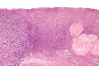
 Ovary with a metastasis from breast carcinoma (left of image). H&E stain. | |
|
| |
| LM | predominantly surface involvement and nodular (especially at low power), +/-signet ring cells (suggestive of GI or breast primary), lymphovascular invasion |
| LM DDx | ovarian primary tumours - esp. ovarian mucinous carcinoma, serous carcinoma of the ovary, endometrioid carcinoma of the ovary |
| IHC | dependent on primary site |
| Gross | classically bilateral masses <10 cm |
| Site | ovary - see ovarian tumours |
|
| |
| Clinical history | +/-malignancy elsewhere |
| Signs | +/-abdominal mass |
| Symptoms | +/-pain, +/-abdominal distension |
| Blood work | serology suggestive of other primary, e.g. elevated CEA |
| Radiology | adnexal mass/ovarian mass |
| Prognosis | poor |
| Clin. DDx | ovarian primary |
| Treatment | dependent on primary - often chemotherapy and radiation, occasionally de-bulking (surgery) |
An ovarian metastasis (also ovary with metastatic disease and metastatic ovarian disease) is cancer in the ovary that arose elsewhere and spread to the ovary.
Generally
- Mostly Muellerian origin (uterus, fallopian tube) or pelvic peritoneum.
Common extramuellerian metastatic tumours:
- Breast.
- Gastrointestinal (GI) tract.
- Pseudomyxoma peritonei, usu. appendiceal origin.
- Krukenberg tumour = signet ring cell cancer with mucin production of GI origin.
Gross
Features favouring metastatic mucinous carcinoma (from the GI tract) over primary ovarian mucinous carcinoma:[1]
- Bilaterality - both ovaries involved.
- Small unilateral tumour size.
- <10 cm suggests metastasis.
- >13 cm = ovarian primary.
Microscopic
Features:
- Predominantly surface involvement and nodular at low power.
- +/-Signet ring cells (suggestive of GI or breast primary). ‡
- Lymphovascular invasion.
Note:
- ‡ Primary signet ring cell carcinoma of the ovary is reported but very rare.[2]
Images
Breast carcinoma
Urothelial carcinoma
IHC
Ovarian tumours:
- Dipeptidase 1 (DPEP1) +ve.[3]
- CK7 +ve.
See also
References
- ↑ Yemelyanova, AV.; Vang, R.; Judson, K.; Wu, LS.; Ronnett, BM. (Jan 2008). "Distinction of primary and metastatic mucinous tumors involving the ovary: analysis of size and laterality data by primary site with reevaluation of an algorithm for tumor classification.". Am J Surg Pathol 32 (1): 128-38. doi:10.1097/PAS.0b013e3180690d2d. PMID 18162780.
- ↑ El-Safadi, S.; Stahl, U.; Tinneberg, HR.; Hackethal, A.; Muenstedt, K. (Sep 2010). "Primary signet ring cell mucinous ovarian carcinoma: a case report and literature review.". Case Rep Oncol 3 (3): 451-7. doi:10.1159/000323003. PMID 21611142.
- ↑ Okamoto, T.; Matsumura, N.; Mandai, M.; Oura, T.; Yamanishi, Y.; Horiuchi, A.; Hamanishi, J.; Baba, T. et al. (Feb 2011). "Distinguishing primary from secondary mucinous ovarian tumors: an algorithm using the novel marker DPEP1.". Mod Pathol 24 (2): 267-76. doi:10.1038/modpathol.2010.204. PMID 21076463.


