Difference between revisions of "Endocervical adenocarcinoma in situ"
Jump to navigation
Jump to search
(→IHC: +SO) |
(→Images: fix) |
||
| Line 78: | Line 78: | ||
Image: Endocervical adenocarcinoma in situ 2b -- very high mag.jpg | AIS - very high mag. | Image: Endocervical adenocarcinoma in situ 2b -- very high mag.jpg | AIS - very high mag. | ||
</gallery> | </gallery> | ||
www | ====www==== | ||
*[ | *[https://www.flickr.com/photos/euthman/1799754415 Endocervical AIS adjacent to normal (flickr.com/euthman)]. | ||
*[http://nih.techriver.net/view.php?patientId=99 Endocervical adenocarcinoma in situ (techriver.net)]. | *[http://nih.techriver.net/view.php?patientId=99 Endocervical adenocarcinoma in situ (techriver.net)]. | ||
*[http://womenshealthsection.com/content/gynpc/gynpc006d.jpg Endocervical adenocarcinoma in situ (womenshealthsection.com)].<ref>URL: [http://www.womenshealthsection.com/content/print.php3?title=gynpc006&cat=60&lng=english http://www.womenshealthsection.com/content/print.php3?title=gynpc006&cat=60&lng=english]. Accessed on: 20 March 2013.</ref> | *[http://womenshealthsection.com/content/gynpc/gynpc006d.jpg Endocervical adenocarcinoma in situ (womenshealthsection.com)].<ref>URL: [http://www.womenshealthsection.com/content/print.php3?title=gynpc006&cat=60&lng=english http://www.womenshealthsection.com/content/print.php3?title=gynpc006&cat=60&lng=english]. Accessed on: 20 March 2013.</ref> | ||
Revision as of 02:35, 18 August 2015
| Endocervical adenocarcinoma in situ | |
|---|---|
| Diagnosis in short | |
|
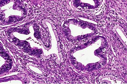
Template:Px Endocervical adenocarcinoma in situ. H&E stain. | |
|
| |
| LM | Nuclear changes (nuclear crowding/pseudostratification, nuclear enlargement (often cigar-shaped nuclei), coarse chromatin, small nucleolus or nucleoli), +/-mitoses, +/-reduced cytoplasmic mucin, preservation of glandular architecture |
| LM DDx | tubal metaplasia, Arias-Stella reaction, endometriosis, lower uterine segment epithelium (esp. proliferative phase endometrium), endocervical adenocarcinoma, metastatic adenocarcinoma |
| IHC | p16 +ve |
| Site | uterine cervix - endocervical canal |
|
| |
| Associated Dx | squamous lesions (LSIL, HSIL) |
| Clinical history | +/-HPV |
| Symptoms | asymptomatic |
| Prevalence | uncommon |
| Clin. DDx | endocervical adenocarcinoma (invasive), endometrial carcinoma, squamous cervical lesions |
| Treatment | typically LEEP |
- For the cytology see Endocervical adenocarcinoma in situ (cytology)
Endocervical adenocarcinoma in situ, also adenocarcinoma in situ of the uterine endocervix, is pre-invasive change of the uterine endocervix. It is closely tied to HPV infection.
If the context is clear, it may be referred to as adenocarcinoma in situ, abbreviated AIS.
General
- Usually due to HPV.
- May be found together with squamous neoplasias of the cervix.
- AIS of the cervix is much less common than squamous dysplasia of the cervix/SCC of the cervix.
- Generally, definitely diagnosed with an endocervical curettage (ECC).
Gross
- Not apparent at colposcopy.
Microscopic
Features:[1]
- Nuclear changes - key feature:
- Variable nuclear stratification.
- Nuclear crowding/pseudostratification.
- Nuclear enlargement.
- Often cigar-shaped nuclei.
- Coarse chromatin.
- Small nucleolus or nucleoli.
- Variable nuclear stratification.
- +/-Mitoses.
- +/-Reduced cytoplasmic mucin.
- Preservation of glandular architecture.
- Normal gland spacing - lack of complexity ("lobular pattern").
- Normal gland depth (subjective).
DDx:
- Tubal metaplasia.
- Arias-Stella reaction.
- Endometriosis.
- Lower uterine segment epithelium[2] - esp. proliferative phase endometrium - mitoses rare, NC ratio normal, stroma different.
- Endocervical adenocarcinoma - often has paradoxical maturation... paler cytoplasm & nuclei than adjacent AIS.
- Metastatic adenocarcinoma.
- Proliferative phase endometrium - endometrial type stroma, cytoplasm not pale staining, no nuclear atypia (smooth nuclear contour, stratified).
Images
- Endocervical adenocarcinoma in situ -- intermed mag.jpg
AIS - intermed. mag.
- Endocervical adenocarcinoma in situ -- high mag.jpg
AIS - high mag.
- Endocervical adenocarcinoma in situ -- very high mag.jpg
AIS - very high mag.
- Endocervical adenocarcinoma in situ 2 -- intermed mag.jpg
AIS - intermed. mag.
- Endocervical adenocarcinoma in situ 2 -- high mag.jpg
AIS - high mag.
- Endocervical adenocarcinoma in situ 2a -- very high mag.jpg
AIS - very high mag.
- Endocervical adenocarcinoma in situ 2b -- very high mag.jpg
AIS - very high mag.
www
- Endocervical AIS adjacent to normal (flickr.com/euthman).
- Endocervical adenocarcinoma in situ (techriver.net).
- Endocervical adenocarcinoma in situ (womenshealthsection.com).[3]
- Endocervical adenocarcinoma in situ - cytology (techriver.net).
IHC
- p16 +ve.
- CEA +ve.
- Vimentin -ve.
Sign out
UTERINE CERVIX, BIOPSY:
- HIGH-GRADE SQUAMOUS INTRAEPITHELIAL LESION (HSIL).
- ENDOCERVICAL ADENOCARCINOMA IN SITU (AIS).
- ACUTE AND CHRONIC INFLAMMATION.

COMMENT:
A p16 immunostain marks the full thickness of the squamous epithelium and is
strong. A Ki-67 immunostain marks increased numbers of superficial squamous cells.

The atypical endocervical epithelium (interpreted as AIS) is strongly p16 positive
and has an increased proliferative activity with Ki-67 staining.
Micro
The atypical endocervical epithelium (interpreted as AIS) shows marked hyperchromasia, nuclear crowding and moderate nuclear atypia with a relatively abundant cytoplasm ( nucleus to cell size = 1:2 ).
See also
References
- ↑ Zaino, RJ. (Mar 2000). "Glandular lesions of the uterine cervix.". Mod Pathol 13 (3): 261-74. doi:10.1038/modpathol.3880047. PMID 10757337. http://www.nature.com/modpathol/journal/v13/n3/full/3880047a.html.
- ↑ Nucci, Marisa R.; Oliva, Esther (2009). Gynecologic Pathology: A Volume in Foundations in Diagnostic Pathology Series (1st ed.). Churchill Livingstone. pp. 167. ISBN 978-0443069208.
- ↑ URL: http://www.womenshealthsection.com/content/print.php3?title=gynpc006&cat=60&lng=english. Accessed on: 20 March 2013.