Difference between revisions of "Papillary craniopharyngioma"
Jump to navigation
Jump to search
(redirect) |
(split-out) |
||
| Line 1: | Line 1: | ||
'''Papillary craniopharyngioma''', abbreviated '''pCP''', is a benign [[brain tumour]] of the sella turcica. | |||
It is a subtype of [[craniopharyngioma]]. | |||
==General== | |||
*Adults individuals.<ref name=pmid6696166>{{Cite journal | last1 = Giangaspero | first1 = F. | last2 = Burger | first2 = PC. | last3 = Osborne | first3 = DR. | last4 = Stein | first4 = RB. | title = Suprasellar papillary squamous epithelioma ("papillary craniopharyngioma"). | journal = Am J Surg Pathol | volume = 8 | issue = 1 | pages = 57-64 | month = Jan | year = 1984 | doi = | PMID = 6696166 }}</ref> | |||
*Usually solid. | |||
==Microscopic== | |||
Features (papillary):<ref name=Ref_PSNP406>{{Ref PSNP|406}}</ref> | |||
*Non-keratinized squamous epithelium (without nuclear atypia). | |||
*Fibrovascular cores (required for ''papillary''). | |||
Notes: | |||
*+/-Cilia (rare). | |||
*+/-Goblet cell-like formations (rare). | |||
===Image=== | |||
<gallery> | |||
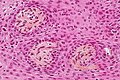
Image: Papillary craniopharyngioma - intermed mag.jpg | PC - intermed. mag. (WC/Nephron) | |||
Image: Papillary craniopharyngioma - high mag.jpg | PC - high mag. (WC/Nephron) | |||
Image: Papillary craniopharyngioma - very high mag.jpg | PC - very high mag. (WC/Nephron) | |||
</gallery> | |||
www: | |||
*[http://library.med.utah.edu/WebPath/jpeg4/ENDO115.jpg Craniopharyngioma (med.utah.edu)].<ref>URL: [http://library.med.utah.edu/WebPath/jpeg4/ENDO115.jpg http://library.med.utah.edu/WebPath/jpeg4/ENDO115.jpg]. Accessed on: 6 December 2010.</ref> | |||
==See also== | |||
*[[Pituitary gland]]. | |||
==References== | |||
{{Reflist|2}} | |||
[[Category:Diagnosis]] | [[Category:Diagnosis]] | ||
Revision as of 01:44, 16 November 2014
Papillary craniopharyngioma, abbreviated pCP, is a benign brain tumour of the sella turcica.
It is a subtype of craniopharyngioma.
General
- Adults individuals.[1]
- Usually solid.
Microscopic
Features (papillary):[2]
- Non-keratinized squamous epithelium (without nuclear atypia).
- Fibrovascular cores (required for papillary).
Notes:
- +/-Cilia (rare).
- +/-Goblet cell-like formations (rare).
Image

PC - intermed. mag. (WC/Nephron)

PC - high mag. (WC/Nephron)
PC - very high mag. (WC/Nephron)


www:
See also
References
- ↑ Giangaspero, F.; Burger, PC.; Osborne, DR.; Stein, RB. (Jan 1984). "Suprasellar papillary squamous epithelioma ("papillary craniopharyngioma").". Am J Surg Pathol 8 (1): 57-64. PMID 6696166.
- ↑ Perry, Arie; Brat, Daniel J. (2010). Practical Surgical Neuropathology: A Diagnostic Approach: A Volume in the Pattern Recognition series (1st ed.). Churchill Livingstone. pp. 406. ISBN 978-0443069826.
- ↑ URL: http://library.med.utah.edu/WebPath/jpeg4/ENDO115.jpg. Accessed on: 6 December 2010.