Difference between revisions of "Glassy cell carcinoma"
Jump to navigation
Jump to search
(split out) |
|||
| (4 intermediate revisions by the same user not shown) | |||
| Line 1: | Line 1: | ||
{{ Infobox diagnosis | |||
| Name = {{PAGENAME}} | |||
| Image = Glassy_cell_carcinoma_-_very_high_mag.jpg | |||
| Width = | |||
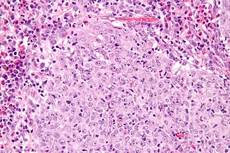
| Caption = Glassy cell carcinoma. [[H&E stain]]. | |||
| Synonyms = | |||
| Micro = epithelioid cells in sheets or cords with a round/oval nucleus, one or more prominent nucleoli, abundant finely vacuolated eosinophilic to amphophilic cytoplasm, distinct cell borders, inflammation - especially eosinophils | |||
| Subtypes = | |||
| LMDDx = [[adenosquamous carcinoma of the uterine cervix]], [[squamous carcinoma of the uterine cervix]] | |||
| Stains = [[PAS stain]] +ve (plasma membrane) | |||
| IHC = | |||
| EM = | |||
| Molecular = | |||
| IF = | |||
| Gross = | |||
| Grossing = | |||
| Site = [[uterine cervix]] | |||
| Assdx = | |||
| Syndromes = | |||
| Clinicalhx = rapid growth | |||
| Signs = | |||
| Symptoms = | |||
| Prevalence = rare | |||
| Bloodwork = | |||
| Rads = | |||
| Endoscopy = | |||
| Prognosis = poor | |||
| Other = | |||
| ClinDDx = other [[uterine cervix|cervical]] tumours | |||
| Tx = | |||
}} | |||
'''Glassy cell carcinoma''', abbreviated '''GCC''', is a rare [[malignancy]] of the [[uterine cervix]]. | '''Glassy cell carcinoma''', abbreviated '''GCC''', is a rare [[malignancy]] of the [[uterine cervix]]. | ||
It is considered to be a subtype of [[adenosquamous carcinoma of the uterine cervix]]. | |||
==General== | ==General== | ||
*Rare. | *Rare. | ||
| Line 8: | Line 40: | ||
==Microscopic== | ==Microscopic== | ||
Features:<ref name=pmid11393075>{{Cite journal | last1 = Reis-Filho | first1 = JS. | last2 = Fillus Neto | first2 = J. | last3 = Schonemann | first3 = E. | last4 = Sanderson | first4 = A. | last5 = Schmitt | first5 = FC. | title = Glassy cell carcinoma of the uterine cervix. Report of a case with cytohistologic and immunohistochemical study. | journal = Acta Cytol | volume = 45 | issue = 3 | pages = 407-10 | month = | year = | doi = | PMID = 11393075 }}</ref> | Features:<ref name=pmid11393075>{{Cite journal | last1 = Reis-Filho | first1 = JS. | last2 = Fillus Neto | first2 = J. | last3 = Schonemann | first3 = E. | last4 = Sanderson | first4 = A. | last5 = Schmitt | first5 = FC. | title = Glassy cell carcinoma of the uterine cervix. Report of a case with cytohistologic and immunohistochemical study. | journal = Acta Cytol | volume = 45 | issue = 3 | pages = 407-10 | month = | year = | doi = | PMID = 11393075 }}</ref> | ||
*Epithelioid cells in sheets or cords | *Epithelioid cells in sheets or cords with: | ||
*Round/oval nucleus. | **Round/oval nucleus. | ||
*One or more prominent nucleoli. | **One or more prominent nucleoli. | ||
*Abundant finely vacuolated eosinophilic to amphophilic cytoplasm. | **Abundant finely vacuolated eosinophilic to amphophilic cytoplasm - looks "glassy". | ||
*Distinct cell borders. | **Distinct cell borders. | ||
*Inflammation - esp. eosinophils.<ref>URL: [http://www.webpathology.com/image.asp?n=2&Case=561 http://www.webpathology.com/image.asp?n=2&Case=561]. Accessed on: 4 September 2011.</ref> | *Inflammation - esp. eosinophils.<ref>URL: [http://www.webpathology.com/image.asp?n=2&Case=561 http://www.webpathology.com/image.asp?n=2&Case=561]. Accessed on: 4 September 2011.</ref> | ||
Latest revision as of 02:40, 25 April 2014
| Glassy cell carcinoma | |
|---|---|
| Diagnosis in short | |
 Glassy cell carcinoma. H&E stain. | |
|
| |
| LM | epithelioid cells in sheets or cords with a round/oval nucleus, one or more prominent nucleoli, abundant finely vacuolated eosinophilic to amphophilic cytoplasm, distinct cell borders, inflammation - especially eosinophils |
| LM DDx | adenosquamous carcinoma of the uterine cervix, squamous carcinoma of the uterine cervix |
| Stains | PAS stain +ve (plasma membrane) |
| Site | uterine cervix |
|
| |
| Clinical history | rapid growth |
| Prevalence | rare |
| Prognosis | poor |
| Clin. DDx | other cervical tumours |
Glassy cell carcinoma, abbreviated GCC, is a rare malignancy of the uterine cervix.
It is considered to be a subtype of adenosquamous carcinoma of the uterine cervix.
General
- Rare.
- Rapid growth, poor prognosis.[1]
- Considered a subtype of adenosquamous carcinoma.[2]
Microscopic
Features:[3]
- Epithelioid cells in sheets or cords with:
- Round/oval nucleus.
- One or more prominent nucleoli.
- Abundant finely vacuolated eosinophilic to amphophilic cytoplasm - looks "glassy".
- Distinct cell borders.
- Inflammation - esp. eosinophils.[4]
DDx:
Images
www:
- GCC - low mag. (webpathology.com).
- GCC - high mag. (webpathology.com).
- GCC - several images (upmc.edu).
Stains
See also
References
- ↑ Nasu, K.; Takai, N.; Narahara, H. (Jun 2009). "Multimodal treatment for glassy cell carcinoma of the uterine cervix.". J Obstet Gynaecol Res 35 (3): 584-7. doi:10.1111/j.1447-0756.2008.00968.x. PMID 19527406.
- ↑ Kosińiska-Kaczyńska, K.; Mazanowska, N.; Bomba-Opoń, D.; Horosz, E.; Marczewska, M.; Wielgoś, M. (Dec 2011). "Glassy cell carcinoma of the cervix--a case report with review of the literature.". Ginekol Pol 82 (12): 936-9. PMID 22384631.
- ↑ Reis-Filho, JS.; Fillus Neto, J.; Schonemann, E.; Sanderson, A.; Schmitt, FC.. "Glassy cell carcinoma of the uterine cervix. Report of a case with cytohistologic and immunohistochemical study.". Acta Cytol 45 (3): 407-10. PMID 11393075.
- ↑ URL: http://www.webpathology.com/image.asp?n=2&Case=561. Accessed on: 4 September 2011.
- ↑ Deshpande, AH.; Kotwal, MN.; Bobhate, SK.. "Glassy cell carcinoma of the uterine cervix a rare histology. Report of three cases with a review of the literature.". Indian J Cancer 41 (2): 92-5. PMID 15318016.


