Difference between revisions of "Sinus histiocytosis"
Jump to navigation
Jump to search
(finish split-out) |
|||
| Line 29: | Line 29: | ||
*[[Lymph node pathology]]. | *[[Lymph node pathology]]. | ||
*[[Dermatopathic lymphadenopathy]] | *[[Dermatopathic lymphadenopathy]] | ||
==References== | |||
{{Reflist|1}} | |||
[[Category:Diagnosis]] | [[Category:Diagnosis]] | ||
[[Category:Lymph node pathology]] | [[Category:Lymph node pathology]] | ||
Revision as of 00:21, 1 December 2013
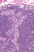
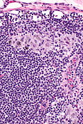
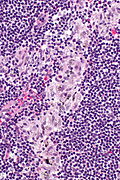
Sinus histiocytosis, abbreviated SH, is a common finding in lymph nodes.
It should not be confused with Rosai-Dorfman disease (also known as sinus histiocytosis and massive lymphadenopathy).
General
- Benign.
- Non-specific finding.
Microscopic
Features:[1]
- Sinuses distended with histiocytes - key feature.
- Plasma cells increased.
DDx:
- Rosai-Dorfman disease - histiocyte nuclei large (~2-3x lymphocyte) and round with a prominent nucleoli.
- Dermatopathic lymphadenopathy - histiocytes have (melanin) pigment.
Images
Sign out
- The finding is often ignored; may be signed out as morphologically benign lymph nodes.
See also
References
- ↑ Ioachim, Harry L; Medeiros, L. Jeffrey (2008). Ioachim's Lymph Node Pathology (4th ed.). Lippincott Williams & Wilkins. pp. 179. ISBN 978-0781775960.


