Difference between revisions of "Bone"
m (→Microscopic: wikify) |
|||
| Line 218: | Line 218: | ||
===Microscopic=== | ===Microscopic=== | ||
*Macrophages in the marrow space with a "crumpled tissue paper" appearance. | *Macrophages in the marrow space with a "crumpled tissue paper" appearance. | ||
==Langerhans cell histiocytosis of bone== | |||
*[[AKA]] ''eosinophilic granuloma of bone''. | |||
{{Main|Langerhans cell histiocytosis}} | |||
===General=== | |||
*Rare. | |||
===Microscopic=== | |||
*Eosinophils. | |||
*Cerebriform and/or reniform macrophages. | |||
=See also= | =See also= | ||
Revision as of 00:29, 28 November 2011
Bone is a scaffold it bears weight and occasionally gets infected.
Tumours often spread to bone and occasionally arise in bone. Bone tumours are dealt with in the bone tumours article.
Normal bone
Bone
Matrix
Two types (based on arrangement of collagen):
- Woven.
- Always abnormal in adults.
- Collagen arranged haphazardly - mechanically weak.
- Lamellar.
- Collagen organized in layers (lamellae).
Woven vs. lamellar:
- Easiest way to differentiate: polarize; lamellar bone has well-defined layers.
Images:
Cells
- Osteocytes.
- Sit in lacunae.
- Empty lacunae = necrotic bone.
- Sit in lacunae.
- Osteoblasts.
- Make bone.
- Osteoclasts.
- Destroy bone.
- Multinucleated.
Memory device: 'b' before 'c'.
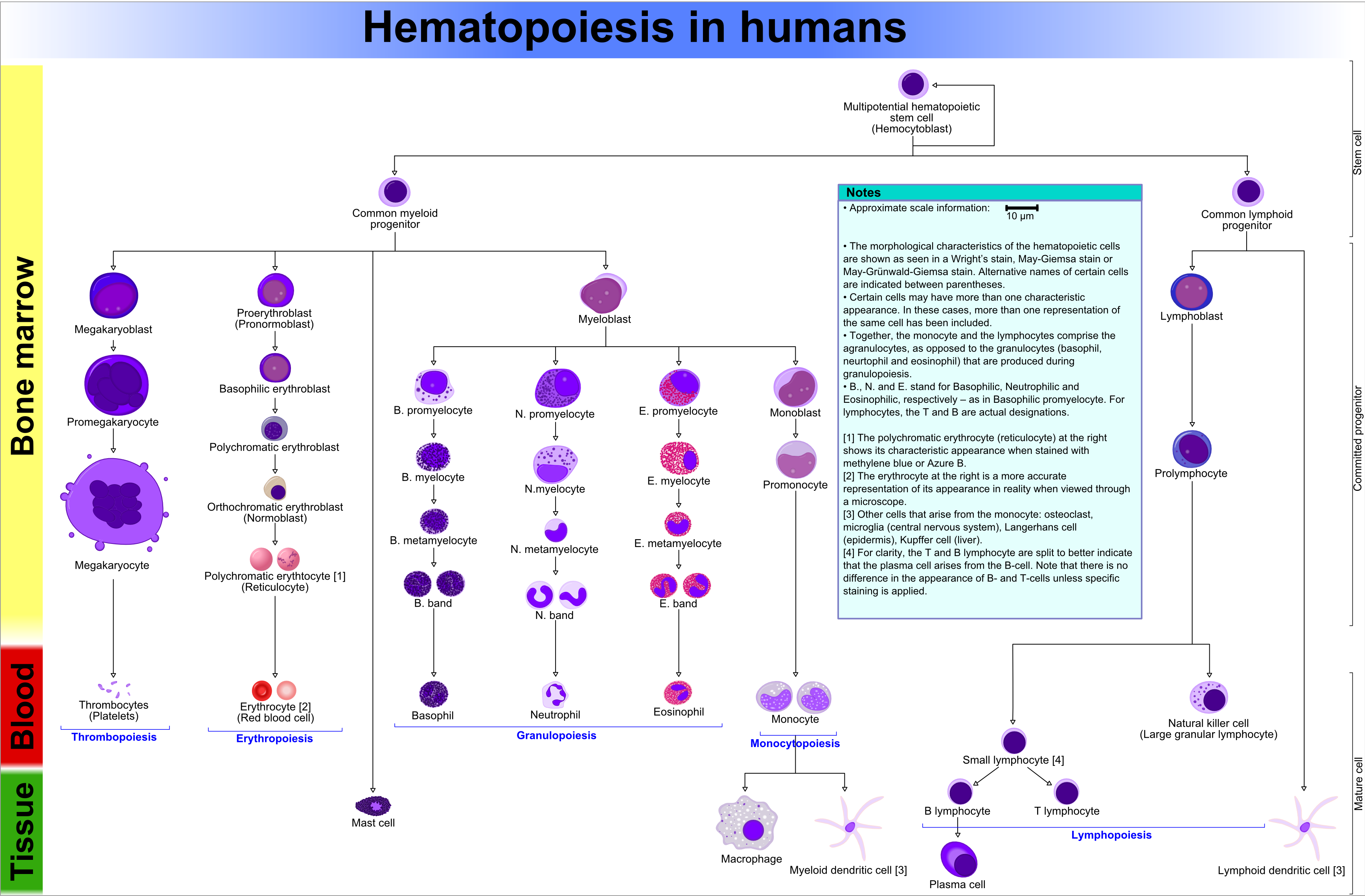
Bone marrow
- Fat content (%) ~= age (in years)[2]
- e.g. 60 year old will have 60% fatty replacement.
- Should see three cell lines.
- The cell lines:[3]
- Erythroid (red cells),
- Myeloid (white blood cells),
- Megakaryocytic (platelets).
- The cell lines:[3]
Note: Lymphocytes are considered separately and typically spared in bone marrow failure.[4]
Identifying the lines:[5]
- Megakaryocytes:
- Big cells ~ 3x the size of a RBC.
- Normoblasts (RBC precursors):
- Hyperchromatic, i.e. blue, nucleus.
- Myeloid line:
- Granules.
- Reniform nucleus, i.e. kidney bean shaped nucleus.
Images:
Organization
- Mature hematopoeitic cells at the centre (distant from bone).
- Immature hematopoeitic cells adjacent to the bone.
Benign variants
Hyperostosis frontalis interna
- Extra-thick frontal bone.[6]
- No clinical significance -- just has to be recognized as a "nothing".
Infections
Osteomyelitis
General
- Hematogenous - often in children.
- Direct entry (skin defect) - adults with diabetes.
Microscopic
- PMNs.
Chronic osteomyelitis
Microscopic
Features:
Bone tumours
This is a big topic. It is dealt with in a separate article.
The bone tumour article covers tumour mimics, e.g. brown cell tumour.
Fractures
This is dealt with in the forensic pathology article.
Others
The following is a collection of stuff that doesn't really fit in another category or is just weird.
A general DDx for cystic bone lesions is found on radipedia.com.[8]
Osteoarthritis
This keeps orthopaedic surgeons busy.
Aneurysmal bone cyst
- Abbreviated ABC.
General
Features:[9]
- Benign.
- May grow rapidly.
- Osteolysis -> cystic space -> filled with blood.
- Relatively common; in children second only to osteosarcoma.[10]
Microscopic
Features:[9]
- Bony trabeculae or osteoid tissue.
- Osteoclast giant cells.
- Multi-nucleated giant-cells with round randomly arranged nuclei.
- Fibroblasts - surround bone.
DDx:
- Giant cell tumour of bone.
- Telangiectatic osteosarcoma.
Myositis ossificans
General
Epidemiology:
- Young people.
- History of trauma - typically.
- Extremities - digits (fingers, toes).
Notes:
- Histologically "worrisome" (for malignancy) - due to high cellularity.[11]
Microscopic
Features:[11]
- High cellularity.
- Low mitotic activity.
- No atypical mitoses.
- No hyperchromasia.
Other features:[12]
- Low power diagnosis:
- Lesion is well-circumscribed.
- Normal muscle is adjacent to the lesion - key feature.
Paget disease of the bone
General
- Benign - unlike Paget disease of the breast.
Classically divided into three phases:[13][14]
- Lytic (predominantly osteoclasts).
- Mixed lytic (osteoclastic) and blastic (osteoblastic).
- Sclerotic (burned-out).
Clinical:
- Elevated ALP.
Microscopic
Features:[13]
- Bone matrix has jigsaw-puzzle like pattern.
- Jigsaw-puzzle pieces each ~ 100-500 micrometres in size (largest dimension).
- Increased osteoclast activity.
- Osteoclast = macrophage that resorbs bone matrix.
Images:
Fibrous dysplasia
- AKA osteitis fibrosa.
General
Classification:
- Monostotic - one bone involved, ~80% of cases.
- Polyostotic - several bones involved, ~20% of cases.
- May be associated with McCune-Albright syndrome.
Microscopic
Features:[15]
- Woven bone with odd irregular shapes - key feature.
- Described as "chinese characters".[16]
- Fibrous tissue around bone.
Notes:
- No osteoblastic rimming.
DDx:
- Desmoplastic fibroma - has lamellar bone.
- Low grade fibrosarcoma.
Image:
- Fibrous dysplasia of bone - high mag. (pathologypics.com).
- Fibrous dysplasia of bone - low mag. (pathologypics.com).
Desmoplastic fibroma
- Not to be confused with desmoplastic fibroblastoma.
General
- Rare.
Microscopic
Features:[17]
- Lamellar bone.
- Fibrotic marrow space with:
- Collagen.
- Low cellularity.
- Spindle cells without significant atypia.
DDx:
- Fibrous dysplasia - has woven bone.
- Low grade fibrosarcoma.
Gaucher disease
General
- May present as a fracture.
Microscopic
- Macrophages in the marrow space with a "crumpled tissue paper" appearance.
Langerhans cell histiocytosis of bone
- AKA eosinophilic granuloma of bone.
General
- Rare.
Microscopic
- Eosinophils.
- Cerebriform and/or reniform macrophages.
See also
- Cartilage.
- Chondro-osseous tumours.
- Femoral head.
- Hematopathology.
- Soft tissue lesions.
- Small round cell tumours.
References
- ↑ Lin DD, Gailloud P, McCarthy EF, Comi AM (February 2006). "Oromaxillofacial osseous abnormality in Sturge-Weber syndrome: case report and review of the literature". AJNR Am J Neuroradiol 27 (2): 274–7. PMID 16484391.
- ↑ IAV. 26 Feb 2009.
- ↑ http://emedicine.medscape.com/article/199003-overview
- ↑ http://emedicine.medscape.com/article/199003-overview
- ↑ http://upload.wikimedia.org/wikipedia/commons/6/69/Hematopoiesis_%28human%29_diagram.png
- ↑ URL: http://radiopaedia.org/articles/hyperostosis_frontalis_interna. Accessed on: 29 September 2010.
- ↑ Alabi, ZO.; Ojo, OS.; Odesanmi, WO. (1991). "Secondary amyloidosis in chronic osteomyelitis.". Int Orthop 15 (1): 21-2. PMID 2071276.
- ↑ URL: http://radipedia.com/WikiMedia/index.php?title=Benign_cystic_bone_lesions. Accessed on: 15 March 2011.
- ↑ 9.0 9.1 URL: http://emedicine.medscape.com/article/1254784-overview. Accessed on: 4 February 2011.
- ↑ van den Berg H, Kroon HM, Slaar A, Hogendoorn P (2008). "Incidence of biopsy-proven bone tumors in children: a report based on the Dutch pathology registration "PALGA"". J Pediatr Orthop 28 (1): 29–35. doi:10.1097/BPO.0b013e3181558cb5. PMID 18157043.
- ↑ 11.0 11.1 11.2 Humphrey, Peter A; Dehner, Louis P; Pfeifer, John D (2008). The Washington Manual of Surgical Pathology (1st ed.). Lippincott Williams & Wilkins. pp. 607. ISBN 978-0781765275.
- ↑ IAV. 9 December 2010.
- ↑ 13.0 13.1 URL: http://emedicine.medscape.com/article/311688-overview. Accessed on: 25 December 2010.
- ↑ URL: http://radiopaedia.org/articles/paget-disease-of-bone-1. Accessed on: 25 December 2010.
- ↑ URL: http://www.pathologypics.com/pictview.aspx?id=104. Accessed on: 14 April 2011.
- ↑ URL: http://www.pathcases.com/bone_tumors_and_tumor.htm. Accessed on: 31 May 2011.
- ↑ URL: http://www.bonetumor.org/tumors-fibrous-tissue/desmoplastic-fibroma. Accessed on: 14 April 2011.